
Electron Microscopy Public Image Archive
Electron Microscopy Public Image Archive
Due to a storage failure, some data files are currently inaccessible (Markted: Incomplete dataset).
We are currently working to restore, but we are accepting priority requests.(email, inquiry).
We are restoring lost files from backups in the following order:
We apologize for the inconvenience and appreciate your understanding.
The EMPIAR-PDBj team at Osaka University assists Asian EM researchers with the transfer of big EM image data to EMPIAR. Instead of sending the data directly to the EBI (UK) via the internet, hard drives can also be sent to Osaka University by postal mail or via a courier service. As an alternative, internet transfer to our server in Osaka is also available. If you would like to take advantage of our submission services, please contact us first by e-mail before sending the data to us.
| Release date | Imageset | Title | Authors and references | Size | Resolution |
|---|---|---|---|---|---|
| 2016-01-27 | 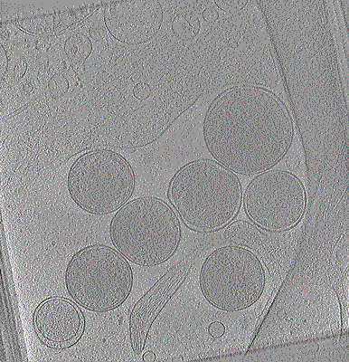 |
Cryo-electron tomogram of Chlamydia trachomatis with type III secretion system in contact with HeLa cell [1 class averages in MRC format] | Nans A, Kudryashev M, Saibil HR, Hayward RD [Pubmed: 26656452] [DOI: 10.1038/ncomms10114] |
13.6 GB | 38.0 Å |
| 2022-05-20 |  |
CEM1.5M : a cellular EM dataset containing ~1.5 x 10⁶ unlabeled 2D image patches curated for deep learning [1592753 micrographs in TIFF format] | Narayan K | 57.6 GB | — |
| 2021-05-28 | 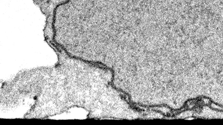 |
CLEM/FIB-SEM Imaging of T Cells after the Formation of Signaling Microclusters at the Immunological Synapse [160 micrographs in TIFF format] | Narayan K | 26.6 MB | — |
| 2022-05-10 |  |
CEM-MitoLab: a dataset of ~22K cellular EM 2D images with label maps of ~135K mitochondrial instances, for deep learning [43720 micrographs in TIFF format] | Narayan K, Conrad RW | 2.8 GB | — |
| 2022-03-15 | 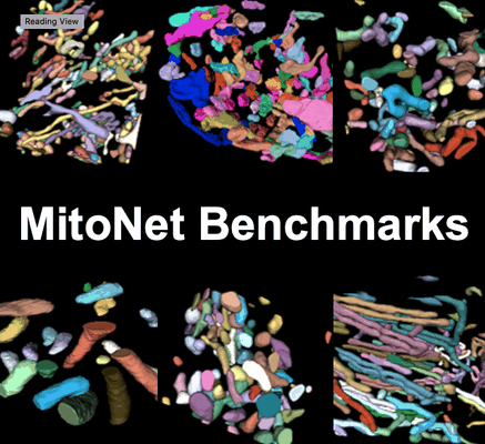 |
Seven benchmark datasets of instance segmentation of mitochondria: 6 diverse volume EM + 1 TEM (100 images) datasets [multiple data sets in TIFF format] | Narayan K, Conrad RW | 4.1 GB | — |
| 2026-08-05 |  |
CryoEM movies of Mo-nitrogenase and FeSII from Azotobacter vinelandii in presence of oxygen [multiple data sets in EER and MRC formats] | Narehood SM, Cook BD, Srisantitham S, Eng VH, Shiau AA, McGuire KL, Britt RD, Herzik Jr MA, Tezcan FA [Pubmed: 39779844] [DOI: 10.1038/s41586-024-08311-1] |
4.9 TB | 2.27 - 3.94 Å |
| 2025-04-20 | 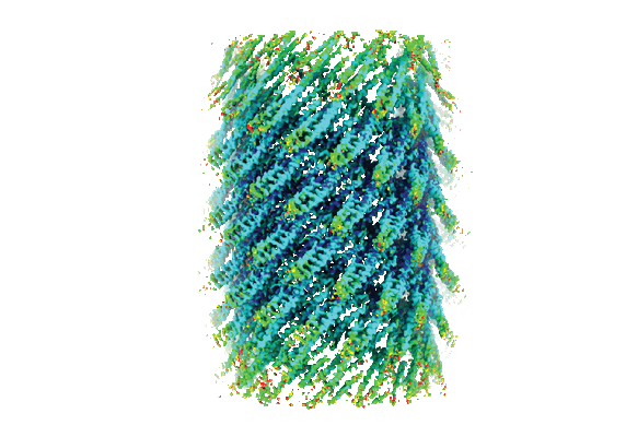 |
Cryo-EM micrographs of Vipp1-F197K/L200K helical filaments (Vipp1-F197K/L200K) [30314 multi-frame micrographs composed of 44 frames each in TIFF format] | Naskar S, Low HH [Pubmed: 39528797] [DOI: 10.1038/s41594-024-01401-8] |
14.1 TB | 3.67 Å |
| 2022-09-20 | 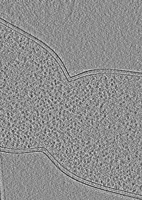 |
Cryo-electron tomography of Cryo-FIB milled dividing E. coli ftsN SPOR domain deletion strain. [60 multi-frame micrographs composed of 4 frames each in MRC format] | Navarro PP, Vettiger A, Ananda VY, Llopis PM, Allolio C, Bernhardt TG, Chao LH [Pubmed: 36097171] [DOI: 10.1038/s41564-022-01210-z] |
189.1 GB | — |
| 2022-09-20 | 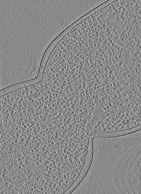 |
Cryo-electron tomography of Cryo-FIB milled dividing E. coli ftsL* strain. [60 multi-frame micrographs composed of 4 frames each in MRC format] | Navarro PP, Vettiger A, Ananda VY, Llopis PM, Allolio C, Bernhardt TG, Chao LH [Pubmed: 36097171] [DOI: 10.1038/s41564-022-01210-z] |
67.2 GB | — |
| 2022-09-20 | 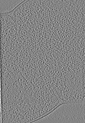 |
Cryo-electron tomography of Cryo-FIB milled dividing E. coli envC and/or nlpD deletion strain. [60 multi-frame micrographs composed of 4 frames each in TIFF format] | Navarro PP, Vettiger A, Ananda VY, Llopis PM, Allolio C, Bernhardt TG, Chao LH [Pubmed: 36097171] [DOI: 10.1038/s41564-022-01210-z] |
73.0 GB | — |
| 2022-09-20 | 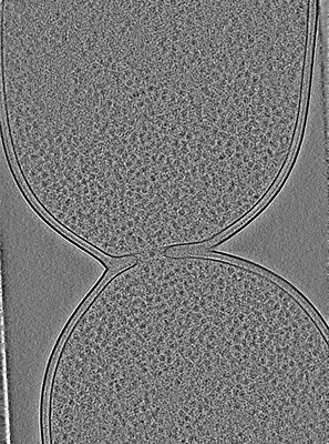 |
Cryo-electron tomography of Cryo-FIB milled dividing E. coli. [60 multi-frame micrographs composed of 4 frames each in TIFF format] | Navarro PP, Vettiger A, Ananda VY, Llopis PM, Allolio C, Bernhardt TG, Chao LH [Pubmed: 36097171] [DOI: 10.1038/s41564-022-01210-z] |
81.5 GB | — |
| 2026-06-28 |  |
Cryo-electron tomography of cryo-FIB milled ∆ponB Escherichia coli division site [61 multi-frame micrographs composed of 4 frames each in TIFF format] | Navarro PP, Vettiger A, Hadju R, Ananda VY, Lopez-Tavares A, Schmid EW, Walter JC, Loose M, Chao LH, Bernhardt TG [Pubmed: 40236067] [DOI: 10.1101/2025.04.02.646830] |
54.4 GB | — |
| 2020-03-23 | 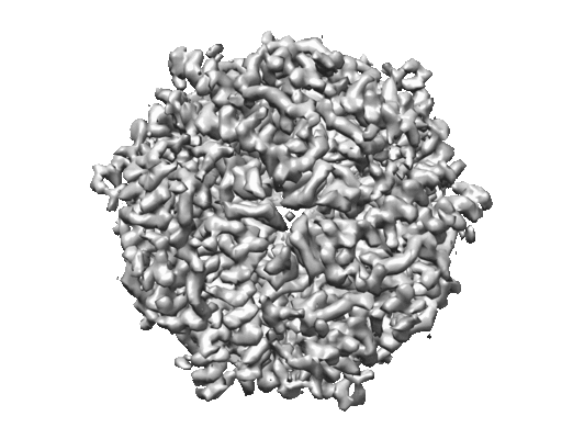 |
Micrographs of DPS collected at 100 keV using a hybrid pixel direct electron detector [739 multi-frame micrographs composed of 32 frames each in MRCS format] | Naydenova K, McMullan G, Peet MJ, Lee Y, Edwards PC, Chen S, Leahy E, Scotcher S, Henderson R, Russo CJ [Pubmed: 31709064] [DOI: 10.1107/S2052252519012612] |
23.3 GB | 3.4 Å |
| 2020-03-23 | 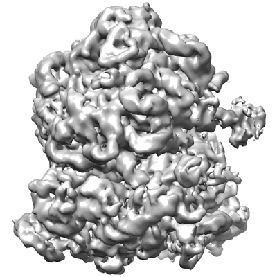 |
Micrographs of E. coli 70S ribosomes collected at 100 keV using a hybrid pixel direct electron detector [127 multi-frame micrographs composed of 32 frames each in MRCS format] | Naydenova K, McMullan G, Peet MJ, Lee Y, Edwards PC, Chen S, Leahy E, Scotcher S, Henderson R, Russo CJ [Pubmed: 31709064] [DOI: 10.1107/S2052252519012612] |
4.0 GB | 7.0 Å |
| 2020-11-27 | 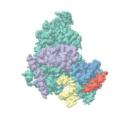 |
Movies of Nsp7-Nsp8-Nsp12 SARS-CoV2 RNA-dependent RNA polymerase in complex with template:primer dsRNA and favipiravir-RTP [63977 multi-frame micrographs composed of 24 frames each in MRCS format] | Naydenova K, Muir KW, Wu LF, Zhang Z, Coscia F, Peet MJ, Castro-Hartmann P, Qian P, Sader K, Dent K, Kimanius D, Sutherland JD, Löwe J, Barford D, Russo CJ [Pubmed: 33526596] [DOI: 10.1073/pnas.2021946118] |
23.9 TB | 2.5 Å |
| 2019-07-29 | 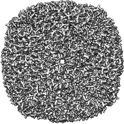 |
Movies of horse spleen apoferritin on multifunctional ultrastable graphene supports for electron cryomicroscopy [480 multi-frame micrographs composed of 38 frames each in MRCS format] | Naydenova K, Peet MJ, Russo CJ [Pubmed: 31127045] [DOI: 10.1073/pnas.1904766116] |
570.0 GB | 2.1 Å |
| 2020-10-21 |  |
Movies of DPS in 260 nm gold foil holes, which eliminate specimen movement [1357 multi-frame micrographs composed of 36 frames each in MRCS format] | Naydenova K, Russo CJ [Pubmed: 33033219] [DOI: 10.1126/science.abb7927] |
691.8 GB | 1.9 Å |
| 2019-10-17 |  |
Cryo electron microscopy of TssA protein from T6SS of Vibrio cholerae. [multiple data sets in MRC and MRCS formats] | Nazarov S, Adaixo R, Basler M [Pubmed: 31403721] [DOI: 10.15252/embj.2018100825] |
1.3 TB | 3.9 Å |
| 2024-11-08 |  |
Cryo-electron microscopy of Avidin [14206 multi-frame micrographs composed of 623 frames each in EER format] | Nazarov S, Beckert Dominique B, Myasnikow A | 3.4 TB | 2.08 Å |
| 2021-11-01 | 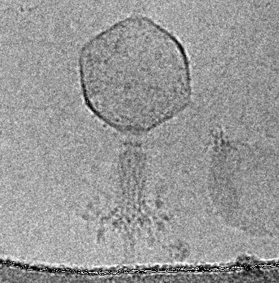 |
The contracted tail of myophage vb_EcoM_CBA120 (CBA120) [535 multi-frame micrographs composed of 20 frames each in MRCS format] | Nazarov S, Leiman P | 674.2 GB | 4.9 Å |
| 2024-03-26 |  |
Cryo-EM structure of mouse heavy-chain apoferritin [9846 multi-frame micrographs composed of 357 frames each in EER format] | Nazarov S.U., Myasnikov A.G., Mohammed I. [Pubmed: 39277607] [DOI: 10.1038/s41467-024-52403-5] |
963.4 GB | 1.09 Å |
| 2022-03-04 |  |
Cryo-electron tomographs of mouse thalamus neurons [60 reconstructed volumes in MRC format] | Nedozralova H, Basnet N, Ibiricu I, Bodakuntla S, Biertümpfel C, Mizuno N [Pubmed: 35262630] [DOI: 10.1083/jcb.202106086] |
71.1 GB | — |
| 2022-03-04 | 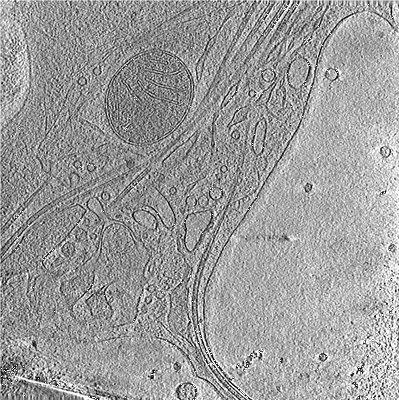 |
Cryo-electron tomographs of mouse hippocampal neurons [63 reconstructed volumes in MRC format] | Nedozralova H, Basnet N, Ibiricu I, Bodakuntla S, Biertümpfel C, Mizuno N [Pubmed: 35262630] [DOI: 10.1083/jcb.202106086] |
88.6 GB | — |
| 2023-02-27 |  |
CryoEM micrographs of mouse apoferritin in a range of ice thicknesses on different microscope setups [multiple data sets in MRC and TIFF formats] | Neselu K, Wang B, Rice WJ, Potter CS, Carragher B, Chua EYD [Pubmed: 36742017] [DOI: 10.1016/j.yjsbx.2023.100085] |
5.7 TB | 2.36 - 10.18 Å |
| 2022-06-07 | 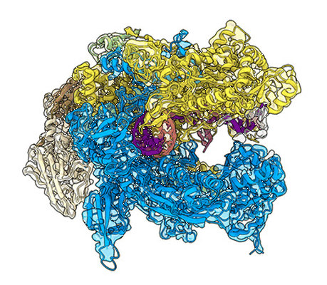 |
Cryo-EM structure of Bacillus subtilis RNA Polymerase elongation complex [4486 multi-frame micrographs composed of 40 frames each in MRC format] | Newing T, Oakley A, Miller M, Dawson C, Brown S, Bouwer J, Tolun G, Lewis P [Pubmed: 33339820] [DOI: 10.1038/s41467-020-20157-5] |
2.3 TB | 3.36 Å |
