Electron Microscopy Public Image Archive
Electron Microscopy Public Image Archive
The EMPIAR-PDBj team at Osaka University assists Asian EM researchers with the transfer of big EM image data to EMPIAR. Instead of sending the data directly to the EBI (UK) via the internet, hard drives can also be sent to Osaka University by postal mail or via a courier service. As an alternative, internet transfer to our server in Osaka is also available. If you would like to take advantage of our submission services, please contact us first by e-mail before sending the data to us.
| Release date | Imageset | Title | Authors and references | Size | Resolution |
|---|---|---|---|---|---|
| 2021-03-12 |  |
PKM2 in complex with L-threonine [476 multi-frame micrographs composed of 75 frames each in TIFF format] | Saur M, Hartshorn MJ, Dong J, Reeks J, Bunkoczi G, Jhoti H, Williams PA [Pubmed: 31877353] [DOI: 10.1016/j.drudis.2019.12.006] |
620.8 GB | 2.6 Å |
| 2021-03-12 |  |
PKM2 in complex with Compound 5 [707 multi-frame micrographs composed of 60 frames each in TIFF format] | Saur M, Hartshorn MJ, Dong J, Reeks J, Bunkoczi G, Jhoti H, Williams PA [Pubmed: 31877353] [DOI: 10.1016/j.drudis.2019.12.006] |
773.8 GB | 3.2 Å |
| 2021-03-12 |  |
PKM2 in complex with Compound 10 [572 multi-frame micrographs composed of 75 frames each in TIFF format] | Saur M, Hartshorn MJ, Dong J, Reeks J, Bunkoczi G, Jhoti H, Williams PA [Pubmed: 31877353] [DOI: 10.1016/j.drudis.2019.12.006] |
745.1 GB | 2.7 Å |
| 2021-03-12 |  |
PKM2 in complex with Compound 6 [590 multi-frame micrographs composed of 75 frames each in TIFF format] | Saur M, Hartshorn MJ, Dong J, Reeks J, Bunkoczi G, Jhoti H, Williams PA [Pubmed: 31877353] [DOI: 10.1016/j.drudis.2019.12.006] |
768.0 GB | 2.5 Å |
| 2023-06-30 | 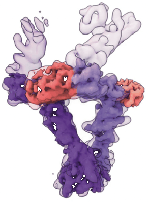 |
CryoEM micrographs of the leptin–leptin receptor complex [multiple data sets in TIFF and MRC formats] | Saxton RA, Caveney NA, Garcia KC [Pubmed: 37002197] [DOI: 10.1038/s41467-023-37169-6] |
11.4 TB | 3.8 - 5.9 Å |
| 2021-04-14 | 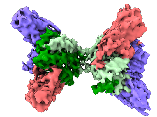 |
Interleukin-10 signaling complex with IL-10RA and IL-10RB [9454 multi-frame micrographs composed of 50 frames each in TIFF format] | Saxton RA, Tsutsumi N, Gati C, Garcia KC [Pubmed: 33737461] [DOI: 10.1126/science.abc8433] |
5.4 TB | 3.5 Å |
| 2021-08-25 | 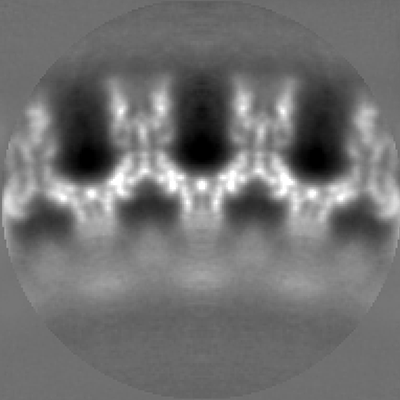 |
Materials for step-by-step guide to efficient subtomogram averaging of virus-like particles with Dynamo [5 reconstructed volumes in MRC format] | Scaramuzza S, Castaño-Díez D [Pubmed: 34437529] [DOI: 10.1371/journal.pbio.3001318] |
645.2 GB | — |
| 2022-11-15 | 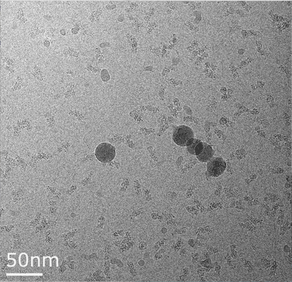 |
cryoEM movies from 30S-RbfA complex [3369 multi-frame micrographs composed of 20 frames each in MRC format] | Schedlbauer A, Fucini P, Connell SR [Pubmed: 34088665] [DOI: 10.1126/sciadv.abf7547] |
3.5 TB | 2.75 - 2.96 Å |
| 2022-11-15 |  |
Movies from 30S-RbfA-RimP-RsmA dataset [4395 multi-frame micrographs composed of 27 frames each in MRCS format] | Schedlbauer A, Fucini P, Connell SR [Pubmed: 34088665] [DOI: 10.1126/sciadv.abf7547] |
3.6 TB | 3.86 - 3.93 Å |
| 2014-11-19 |  |
Beta-galactosidase Falcon-II micrographs plus manually selected coordinates by Richard Henderson [84 micrographs in MRC format] | Scheres SH [Pubmed: 25486611] [DOI: 10.1016/j.jsb.2014.11.010] |
5.3 GB | 4.2 Å |
| 2018-02-26 |  |
Cryo-EM reconstruction of a modified human Adenovirus C5 [231 multi-frame micrographs composed of 50 frames each in MRC format] | Schmid M, Ernst P, Honegger A, Suomalainen M, Zimmermann M, Braun L, Stauffer S, Thom C, Dreier B, Eibauer M, Kipar A, Vogel V, Greber UF, Medalia O, Plückthun A [Pubmed: 29386504] [DOI: 10.1038/s41467-017-02707-6] |
612.7 GB | 7.4 Å |
| 2019-07-29 |  |
CryoWriter: 3.5 Å structure of human 20S proteasome with bound Fabs from microfluidic protein isolation, and 1.9 Å TMV structure [523 multi-frame micrographs composed of 30 frames each in TIFF format] | Schmidli C, Albiez S, Rima L, Righetto R, Mohammed I, Oliva P, Kovacik L, Stahlberg H, Braun T [Pubmed: 31292253] [DOI: 10.1073/pnas.1907214116] |
136.5 GB | 3.5 Å |
| 2024-01-23 | 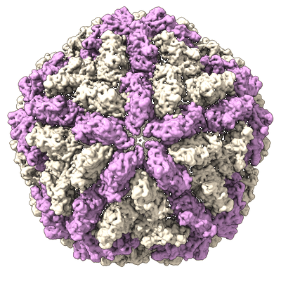 |
Cryo electron microscopy micrographs of high molecular weight fractions from yeast native cell extracts [multiple data sets in MRC format] | Schmidt L, Tueting C, Kyrilis F, Hamdi F, Semchonok DA, Kastritis PL [DOI: 10.1101/2022.07.15.498668] |
4.7 TB | 3.78 - 8.1 Å |
| 2022-04-25 | 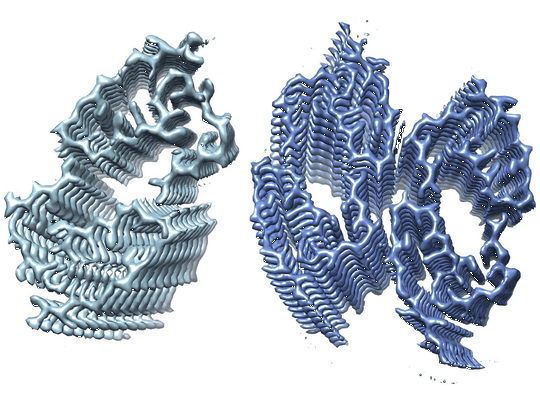 |
Cryo electron microscopy of in vitro recombinant SAA1.1 amyloid fibrils [multiple data sets in TIFF and JPEG formats] | Schmidt MS [Pubmed: 33579941] [DOI: 10.1038/s41467-021-21129-z] |
525.4 GB | 2.73 - 2.95 Å |
| 2021-09-01 | 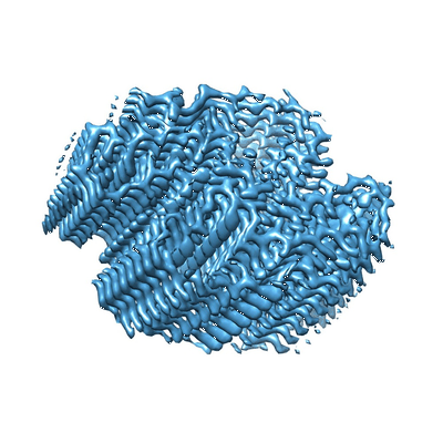 |
Cryo electron microscopy of ex-vivo human SAA amyloid fibrils [6465 multi-frame micrographs composed of 40 frames each in TIFF format] | Schmidt MS [Pubmed: 30846696] [DOI: 10.1038/s41467-019-09033-z] |
1.1 TB | 2.7 Å |
| 2021-11-02 | 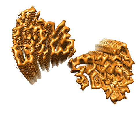 |
Cryo electron microscopy of ex-vivo murine SAA amyloid fibrils [1429 multi-frame micrographs composed of 40 frames each in TIFF format] | Schmidt MS, Fändrich MF [Pubmed: 30846696] [DOI: 10.1038/s41467-019-09033-z] |
533.9 GB | 3.0 Å |
| 2020-09-11 |  |
Spatial Intra- and Intercellular Alignment of Respiratory Cilia and its Relation to Function [16 multi-frame micrographs composed of 100 frames each in MRC format] | Schneiter M, Halm S, Odriozola A, Mogel H, Rička J, Stoffel MH, Zuber B, Frenz M, Tschanz SA [DOI: 10.1101/735332] |
67.5 GB | — |
| 2021-10-18 |  |
Cryo electron tomography of FIB-milled lamella of human DLD-1 cells [3 tilt series in MRC format] | Schuller AP, Wojtynek M, Mankus D, Tatli M, Kronenberg-Tenga R, Regmi SG, Dip PV, Lytton-Jean AKR, Brignole EJ, Dasso M, Weis K, Medalia O, Schwartz TU [Pubmed: 34646014] [DOI: 10.1038/s41586-021-03985-3] |
8.0 GB | 33.0 - 39.0 Å |
| 2022-12-06 |  |
Micrographs of ex vivo AA amyloid from renal tissue of a short hair cat deceased in a shelter [multiple data sets in TIFF format] | Schulte T, Chaves-Sanjuan A, Ricagno S [Pubmed: 36396658] [DOI: 10.1038/s41467-022-34743-2] |
2.0 TB | 3.3 Å |
| 2018-03-21 |  |
Cryo-electron tomography of immature HIV-1 dMACANC VLPs [43 multi-frame micrographs composed of 8 frames each in MRC format] | Schur FK, Obr M, Hagen WJ, Wan W, Jakobi AJ, Kirkpatrick JM, Sachse C, Krausslich HG, Briggs JA, Turoňová B [Pubmed: 27417497] [DOI: 10.1126/science.aaf9620] |
865.0 GB | 3.4 - 3.9 Å |
| 2015-10-09 | 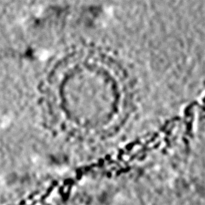 |
Cryo-electron tomography and subtomogram averaging of Rous-Sarcoma-Virus deltaMBD virus-like particles [1 class averages in MRC format] | Schur FKM, Dick RA, Hagen WJH, Vogt VM, Briggs JAG [Pubmed: 26223638] [DOI: 10.1128/JVI.01502-15] |
248.1 MB | — |
| 2022-03-11 |  |
Cryo-EM reconstruction of TMEM106B fold I-d filaments from case 18 with multiple system atrophy [5697 multi-frame micrographs composed of 54 frames each in TIFF format] | Schweighauser M, Arseni D, Bacioglu M, Huang M, Lövestam S, Shi Y, Yang Y, Zhang W, Kotecha A, Garringer HJ, Vidal R, Hallinan GI, Newell KL, Tarutani A, Murayama S, Miyazaki M, Saito Y, Yoshida M, Hasegawa K, Lashley T, Revesz T, Kovacs GG, van Swieten J, Takao M, Hasegawa M, Ghetti B, Spillantini MG, Ryskeldi-Falcon B, Murzin AG, Goedert M, Scheres SHW [Pubmed: 35344985] [DOI: 10.1038/s41586-022-04650-z] |
1.0 TB | 3.64 Å |
| 2020-08-17 |  |
CryoEM dataset of sarkosyl-insoluble fractions from the putamen of multiple system atrophy brain of case 1 [multiple data sets in TIFF, MRC and MRCS formats] | Schweighauser M, Shi Y, Tarutani A, Kametani F, Murzin AG, Ghetti B, Matsubara T, Tomita T, Ando T, Hasegawa K, Murayama S, Yoshida M, Hasegawa M, Scheres SHW, Goedert M [Pubmed: 32461689] [DOI: 10.1038/s41586-020-2317-6] |
3.2 TB | 2.6 Å |
| 2020-11-18 |  |
CryoEM dataset of sarkosyl-insoluble fractions from the putamen of multiple system atrophy brain of case 2 [multiple data sets in TIFF, MRC and MRCS formats] | Schweighauser M, Shi Y, Tarutani A, Kametani F, Murzin AG, Ghetti B, Matsubara T, Tomita T, Ando T, Hasegawa K, Murayama S, Yoshida M, Hasegawa M, Scheres SHW, Goedert M [Pubmed: 32461689] [DOI: 10.1038/s41586-020-2317-6] |
1.1 TB | 3.09 - 3.29 Å |
