
Electron Microscopy Public Image Archive
Electron Microscopy Public Image Archive
Due to a storage failure, some data files are currently inaccessible (Markted: Incomplete dataset).
We are currently working to restore, but we are accepting priority requests.(email, inquiry).
We are restoring lost files from backups in the following order:
We apologize for the inconvenience and appreciate your understanding.
The EMPIAR-PDBj team at Osaka University assists Asian EM researchers with the transfer of big EM image data to EMPIAR. Instead of sending the data directly to the EBI (UK) via the internet, hard drives can also be sent to Osaka University by postal mail or via a courier service. As an alternative, internet transfer to our server in Osaka is also available. If you would like to take advantage of our submission services, please contact us first by e-mail before sending the data to us.
| Release date | Imageset | Title | Authors and references | Size | Resolution |
|---|---|---|---|---|---|
| 2022-09-26 | 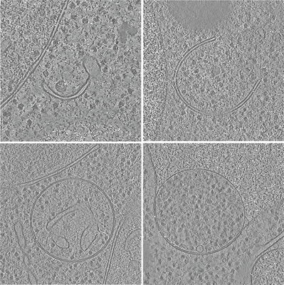 |
In situ cryo-electron tomography of autophagic structures in S. cerevisiae [84 tilt series in MRC format] | Bieber A, Capitanio C, Erdmann PS, Schulman BA, Baumeister W, Wilfling F [Pubmed: 36122245] [DOI: 10.1073/pnas.2209823119] |
249.0 GB | — |
| 2024-09-23 | 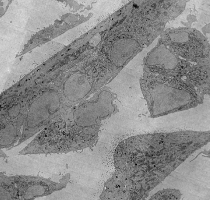 |
FAST-EM array tomography data of wild-type MCF-7 cells prepared with the rOTO protocol and stained with uranyl acetate [5595 micrographs in TIFF format] | Kievits AJ, Duinkerken BHP, Lane R, de Heus C, van Beijeren Bergen en Henegouwen D, Höppener T, Wolters AHG, Liv N, Giepmans BNG, Hoogenboom JP | 561.3 GB | — |
| 2017-12-18 | 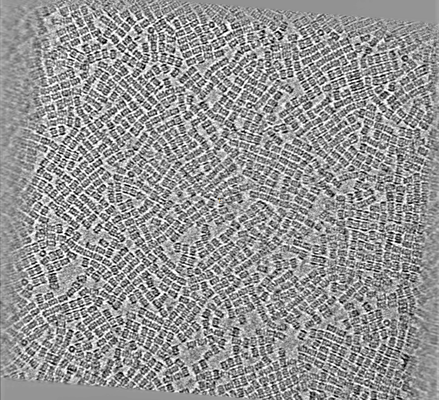 |
CryoET of T20S proteasome single particle [multiple data sets in MRC format] | Noble AJ, Dandey VP, Wei H, Brasch J, Chase J, Acharya P, Tan YZ, Zhang Z, Kim LY, Scapin G, Rapp M, Eng ET, Rice WJ, Cheng A, Negro CJ, Shapiro L, Kwong PD, Jeruzalmi D, des Georges A, Potter CS, Carragher B [Pubmed: 29809143] [DOI: 10.7554/eLife.34257] |
235.3 GB | — |
| 2017-12-18 | 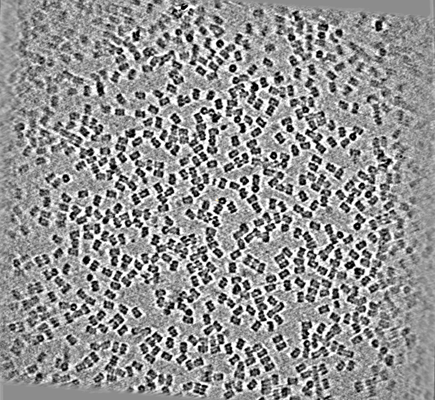 |
CryoET of T20S proteasome single particle [multiple data sets in MRC format] | Dandey VP, Wei H, Brasch J, Chase J, Acharya P, Tan YZ, Zhang Z, Kim LY, Scapin G, Rapp M, Eng ET, Rice MJ, Cheng A, Negro CJ, Shapiro L, Kwong PD, Jeruzalmi D, des Georges A, Potter CS, Carragher B [Pubmed: 29809143] [DOI: 10.7554/eLife.34257] |
12.4 GB | — |
| 2025-06-11 | 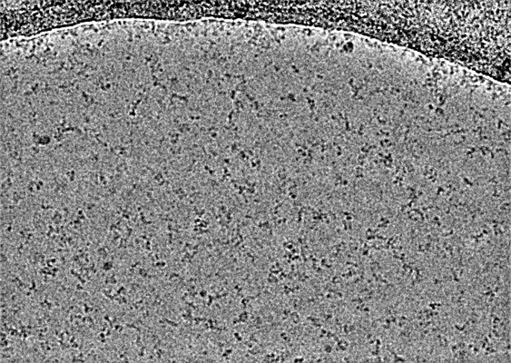 |
Raw data used to generate the structure of the MIRO/TRAK complex [multiple data sets in TIFF format] | Baltrusaitis EE, Ravitch EE, Barrie KR, Dominguez R | 6.9 TB | — |
| 2017-12-18 | 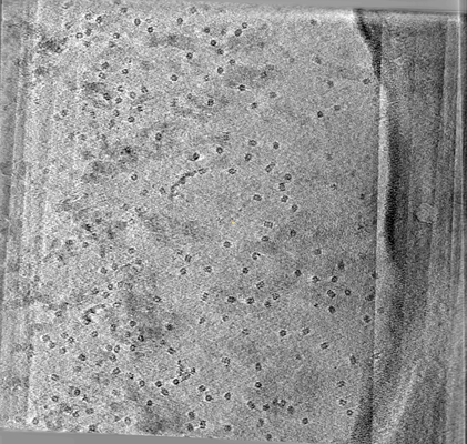 |
CryoET of Mtb 20S proteasome single particle [multiple data sets in MRC format] | Noble AJ, Dandey VP, Wei H, Brasch J, Chase J, Acharya P, Tan YZ, Zhang Z, Kim LY, Scapin G, Rapp M, Eng ET, Rice MJ, Cheng A, Negro CJ, Shapiro L, Kwong PD, Jeruzalmi D, des Georges A, Potter CS, Carragher B [Pubmed: 29809143] [DOI: 10.7554/eLife.34257] |
9.1 GB | — |
| 2024-11-19 |  |
FAST-EM array tomography data of rat pancreas prepared with rOTO protocol and stained with neodymium acetate [4805 micrographs in TIFF format] | Kievits AJ, Duinkerken BHP, Lane R, de Heus C, van Beijeren Bergen en Henegouwen D, Höppener T, Wolters AHG, Liv N, Giepmans BNG, Hoogenboom JP [Pubmed: 39119255] [DOI: 10.1515/mim-2024-0005] |
479.6 GB | — |
| 2018-03-09 | 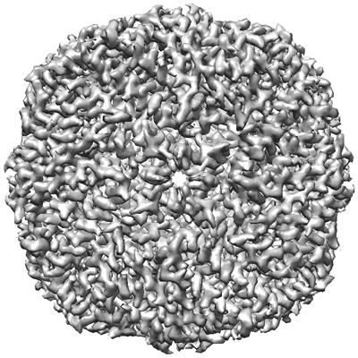 |
Apoferritin tutorial dataset for cisTEM [20 multi-frame micrographs composed of 50 frames each in MRC format] | Grant T, Rohou A, Grigorieff N [Pubmed: 29513216] [DOI: 10.7554/elife.35383] |
5.5 GB | — |
| 2018-01-03 | 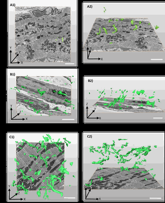 |
3D reconstruction of the tubular system of preterm G55/57, mid term G58/60 and term G66/68 of guinea pig left ventricles [318 multi-frame micrographs composed of 1 frames each in TIFF format] | Kashbour H, Taggart M | 2.9 GB | — |
| 2023-10-17 | 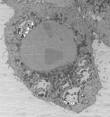 |
3D reconstructions of parasite development and the intracellular niche of the microsporidian pathogen E. intestinalis [multiple data sets in DM4 format] | Antao NVA, Lam CKL, Davydov AD, Riggi MR, Sall JS, Petzold CP, Liang FL, Iwasa JI, Ekiert DCE, Bhabha GB [Pubmed: 37425741] [DOI: 10.1101/2023.07.02.547383] |
537.9 GB | — |
| 2024-11-19 |  |
Training Set for CryoVesNet - Cryo-electron tomograms of rat synaptosomes and synaptic vesicle segmentation [multiple data sets in MRC format] | Khosrozadeh A, Seeger R, Witz G, Radecke J, Sørensen JB, Zuber B [Pubmed: 36876590] [DOI: 10.1101/2022.04.10.487799] |
8.9 GB | — |
| 2018-01-03 | 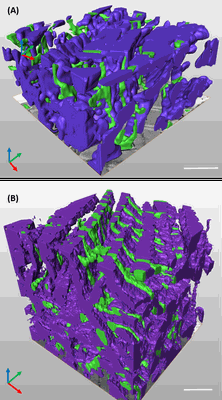 |
3D reconstruction of the cardiac mitochondria in Neaonate and Adult GP cardiomyocyte [262 multi-frame micrographs composed of 1 frames each in TIFF format] | Kashbour H, Taggart M, White K | 1.2 GB | — |
| 2020-05-01 | 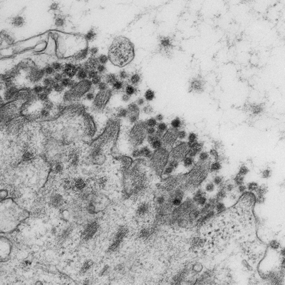 |
SARS-CoV-2 productively infects human gut enterocytes [multiple data sets in TIFF format] | Lamers MM, Beumer J, van der Vaart J, Knoops K, Puschhof J, Breugem T, Ravelli RBG, van Schayck JP, Mykytyn AZ, Duimel HQ, van Donselaar E, Riesebosch S, Kuijpers HJH, Schipper D, van de Wetering WJ, de Graaf M, Koopmans M, Cuppen E, Peters PJ, Haagmans B, Clevers H [Pubmed: 32358202] [DOI: 10.1126/science.abc1669] |
156.2 GB | — |
| 2018-01-03 | 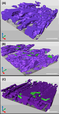 |
3D reconstruction of cardiac mitochondria in earlly prenatal (G55/57) and mid term(G58/60) and term (G66/68) GP cardiomyocyte [341 multi-frame micrographs composed of 1 frames each in TIFF format] | Kashbour H, Taggart M, White K | 1.7 GB | — |
| 2018-01-03 | 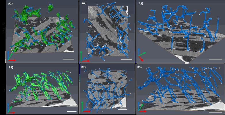 |
Segment /nodes network analysis of the tubular system of the Neonate and Adult GP cardiomyocyte [167 multi-frame micrographs composed of 1 frames each in TIFF format] | Kashbour H, Taggart M, White K | 787.5 MB | — |
| 2025-01-30 | 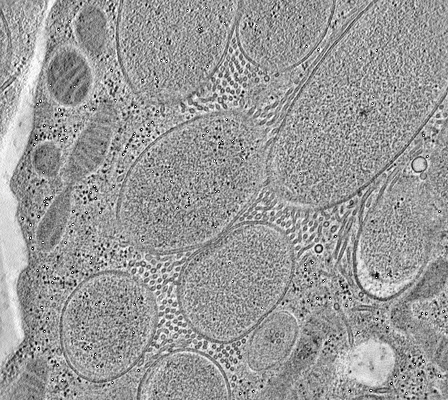 |
Electron tomography of endosymbiotic bacteria inside a bacteriocyte of the cereal weevil Sitophilus oryzae [120 reconstructed volumes in MRC format] | Santarella-Mellwig RSM, Balmand SB, Schwab YS, Zaidman-Remy AZR | 1.0 GB | — |
| 2018-01-03 | 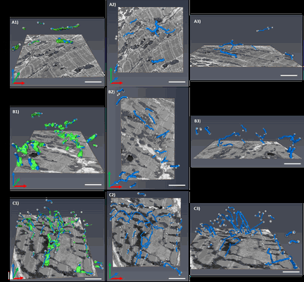 |
Segment /nodes network analysis of the tubular system of the prenatal stages of GP cardiomyocyte [254 multi-frame micrographs composed of 1 frames each in TIFF format] | Kashbour H, Taggart M | 1.9 GB | — |
| 2023-10-03 | 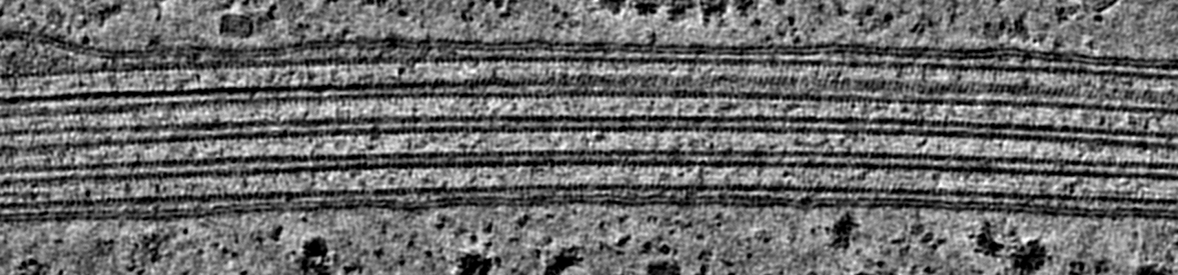 |
Cryo electron tomography of Cytochalasin D-induced protrusions of Drosophila S2 cells treated with thapsigargin or MG132 [multiple data sets in TIFF and MRC formats] | Ventura Santos C, Carter AP [Pubmed: 37702953] [DOI: 10.15252/embr.202357264] |
107.6 GB | — |
| 2025-09-15 |  |
Serial Block Face - Scanning Electron Microscopy of brain microvessels with/without P. falciparum egress products [multiple data sets in TIFF format] | Long RKML, Ronchi PR, Bernabeu MB [DOI: 10.1101/2024.03.29.587334] |
2.3 GB | — |
| 2026-10-05 |  |
2026 CAHRA Challenge 3) Pose Entanglement [stack of 30000 particles in MRCS format] | Feathers JR, Zhong ED, Heeter R, Woollard G, Hanson SM, Cossio P, Greer JAT, Burnley T | 7.3 GB | — |
| 2018-01-17 | 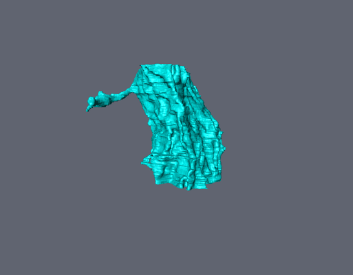 |
Serial Block Face Scanning Electron Micrscopy dataset of the optic lobe from an adult locust [100 micrographs in TIFF format] | Cocks ET [DOI: 10.1111/jmi.12676] |
3.4 GB | — |
| 2025-08-01 |  |
Dataset 3: 70S ribosome with 3 GE81112 bound (Complex 5) [19615 multi-frame micrographs composed of 40 frames each in TIFF format] | Schedlbauer A, Han X, van Bakel W, Kaminishi T, Ochoa-Lizarralde B, Iturrioz I, Çapuni R, Parry R, Zegarra R, Gil-Carton D, López-Alonso JP, Sanz KB, Brandi L, Gualerzi CO, Fucini P, Connell SR [DOI: 10.1101/2024.09.26.614503] |
3.7 TB | — |
| 2026-04-27 |  |
Time-resolved cryo-EM dataset of insulin degrading enzyme in the presence of a 5x molar excess of insulin with a 123 ms freeze time [10932 multi-frame micrographs composed of 50 frames each in TIFF format] | Tang WJ, Mancl JM [DOI: 10.7554/eLife.105761.1] |
5.3 TB | — |
| 2026-09-03 |  |
Cryo-electron tomography of functionally validated synapses in human induced neurons [multiple data sets in MRC format] | Sun RS | 164.1 GB | — |
| 2022-03-04 | 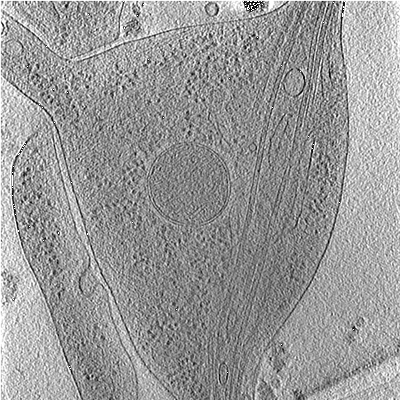 |
Cryo-electron tomographs of mouse thalamus neurons [60 reconstructed volumes in MRC format] | Nedozralova H, Basnet N, Ibiricu I, Bodakuntla S, Biertümpfel C, Mizuno N [Pubmed: 35262630] [DOI: 10.1083/jcb.202106086] |
71.1 GB | — |
