
Electron Microscopy Public Image Archive
Electron Microscopy Public Image Archive
Currently, due to a storage failure, some datasets are either inaccessible or incomplete (Marked: Incomplete dataset).
We are restoring them in ascending order of EMPIAR-ID.
If there are any entries mirrored on PDBj that are particularly urgent, we are accepting requests (email, inquiry).
The EMPIAR-PDBj team at Osaka University assists Asian EM researchers with the transfer of big EM image data to EMPIAR. Instead of sending the data directly to the EBI (UK) via the internet, hard drives can also be sent to Osaka University by postal mail or via a courier service. As an alternative, internet transfer to our server in Osaka is also available. If you would like to take advantage of our submission services, please contact us first by e-mail before sending the data to us.
| Release date | Imageset | Title | Authors and references | Size | Resolution |
|---|---|---|---|---|---|
| 2022-01-24 | 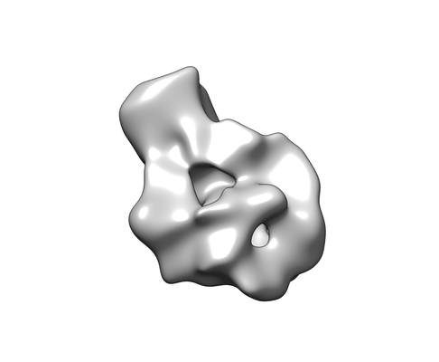 |
Structural Insights of Transcriptionally Active, Full-Length Androgen Receptor Coactivator Complexes [multiple data sets in MRC and TIFF formats] | Yu X, Yi P [Pubmed: 32668201] [DOI: 10.1016/j.molcel.2020.06.031] |
2.0 TB | 20.0 Å |
| 2024-10-08 | 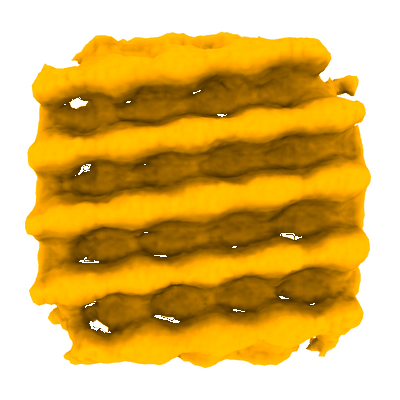 |
In situ cryo-electron tomography of the T. pseudonana diatom PyShell [multiple data sets in MRC and TIFF formats] | Demulder M, Righetto RD, Wietrzynski W, Lamm L, Engel BD [Pubmed: 39357521] [DOI: 10.1016/j.cell.2024.09.013] |
81.8 GB | 20.0 Å |
| 2021-07-07 |  |
Locating Macromolecular Assemblies in Cells by 2D Template Matching with cisTEM [multiple data sets in MRC format] | Lucas BA, Himes BA, Xue L, Grant T, Mahamid J, Grigorieff N [Pubmed: 34114559] [DOI: 10.7554/eLife.68946] |
11.0 GB | 20.0 Å |
| 2024-11-21 |  |
Cryo electron tomography of Echovirus 30-infected cos-7 cells [6 tilt series in MRC format] | Ishemgulova A, Mukhamedova L, Trebichalská Z, Füzik T [Pubmed: 30850609] [DOI: 10.1038/s41467-019-09132-x] |
44.7 GB | 19.4 Å |
| 2022-01-07 | 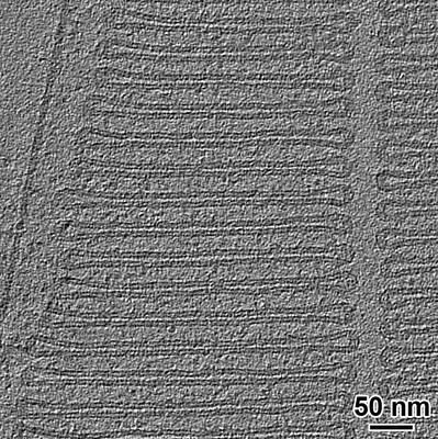 |
Cryo-electron tomography of rod outer segments in wild type mice acquired conventionally with defocus [2 reconstructed volumes in MRC format] | Poege M, Mahamid J, Imanishi SS, Plitzko JM, Palczewski K, Baumeister W [Pubmed: 34931611] [DOI: 10.7554/eLife.72817] |
4.1 GB | 19.3 Å |
| 2023-09-12 | 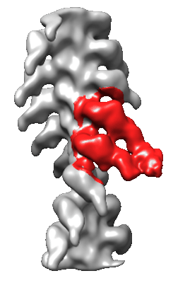 |
CryoEM structure determination of actin decorated with smooth muscle heavy meromyosin [1507 multi-frame micrographs composed of 23 frames each in MRC format] | Hojjatian AH, Liu JL, Taylor DWT, Daneshparvar ND, Winkler HW, Ye FY, Fagnant PMF, Trybus KMT, Taylor KAT [Pubmed: 37414375] [DOI: 10.1016/j.jsb.2023.107995] |
4.2 TB | 19.0 Å |
| 2023-08-18 | 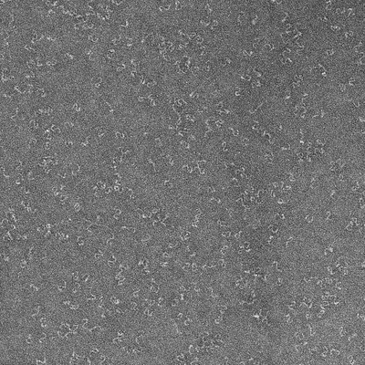 |
Negative stain EM structure of the NF155 extracellular domain [95 micrographs in MRC format] | McKie SJ, Deane JE [Pubmed: 36996106] [DOI: 10.1073/pnas.2218823120] |
1.5 GB | 19.0 Å |
| 2026-10-26 |  |
Split Ring DNA Origami Structure [1116 multi-frame micrographs composed of 61 frames each in MRC format] | Heikkilä ES, Kipnis A, Lautamatti T, Natarajan AK, Kuzyk A | 4.2 TB | 19.0 Å |
| 2023-06-20 | 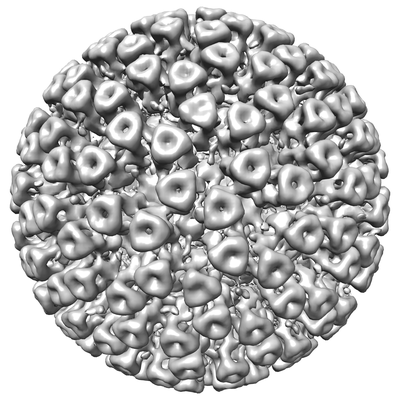 |
Cryo-EPty SPA at CSA of 4.83 mrad [22 micrographs in MRC format] | Pei X, Zhou L, Huang C, Boyce M, Kim JS, Liberti E, Hu Y, Sasaki T, Nellist PD, Zhang P, Stuart DI, Kirkland AI, Wang P [Pubmed: 37230988] [DOI: 10.1038/s41467-023-38268-0] |
3.9 GB | 18.6 Å |
| 2025-03-31 |  |
CryoEM structural characterization of α-Synuclein Oligomer [stack of 2528 particles in MRCS format] | Santos J, Cuellar J, Pallarès I, Valpuesta JM, Ventura S [Pubmed: 38683963] [DOI: 10.1021/jacs.4c02262] |
37.3 GB | 18.3 Å |
| 2025-04-09 | 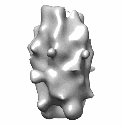 |
CryoEM structural characterization of α-Synuclein Oligomers in complex with PSMa3 peptide [stack of 135393 particles in MRCS format] | Santos J, Cuellar J, Pallarès I, Valpuesta JM, Ventura S [Pubmed: 38683963] [DOI: 10.1021/jacs.4c02262] |
17.8 GB | 18.3 Å |
| 2021-11-08 | 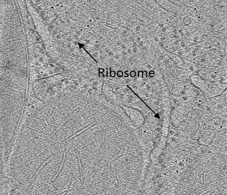 |
VHUT-cryo-FIB, a method to fabricate frozen-hydrated lamella of tissue specimen for in situ cryo-electron tomography [13 multi-frame micrographs composed of 30 frames each in TIFF format] | Zhang J [Pubmed: 34174447] [DOI: 10.1016/j.jsb.2021.107763] |
112.1 GB | 18.0 Å |
| 2020-09-11 | 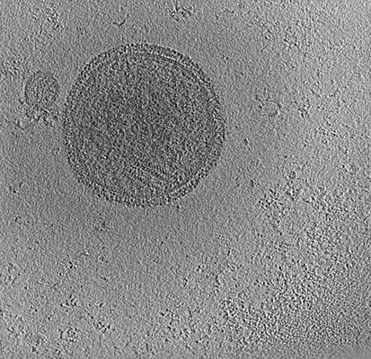 |
Human Parainfluenza Virus Fusion Complex Glycoproteins Imaged In Action On Authentic Viral Surfaces [2 tilt series in MRC format] | Marcink TC, Wang T, des Georges A, Porotto M, Moscona A [Pubmed: 32956394] [DOI: 10.1371/journal.ppat.1008883] |
11.6 GB | 17.18 Å |
| 2024-06-05 | 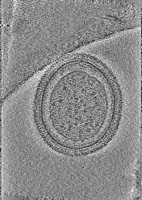 |
Native structure of a human GBP defense complex for cell-autonomous immunity to infection [280 tilt series in TIFF format] | MacMicking JDM, Zhu SZ [Pubmed: 38422126] [DOI: 10.1126/science.abm9903] |
9.7 GB | 17.0 Å |
| 2022-07-12 | 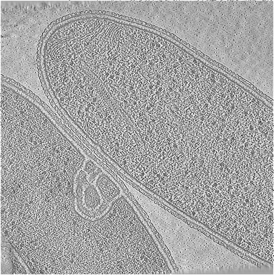 |
In situ cryo-electron tomography of T. kivui cells [multiple data sets in MRC and TIFF formats] | Dietrich HM, Righetto RD, Kumar A, Wietrzynski W, Trischler R, Schuller SK, Wagner J, Schwarz FM, Engel BD, Müller V, Schuller JM [Pubmed: 35859174] [DOI: 10.1038/s41586-022-04971-z] |
373.4 GB | 17.0 Å |
| 2021-01-22 | 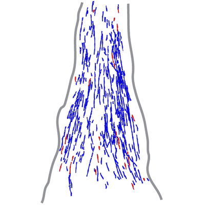 |
Actin filament structure from focal adhesions of mouse embryonic fibroblasts [7 tilt series in MRC format] | Martins B, Sorrentino S, Chung WL, Tatli M, Medalia O, Eibauer M [Pubmed: 33476550] [DOI: 10.1016/j.str.2020.12.014] |
314.1 GB | 17.0 Å |
| 2023-02-28 | 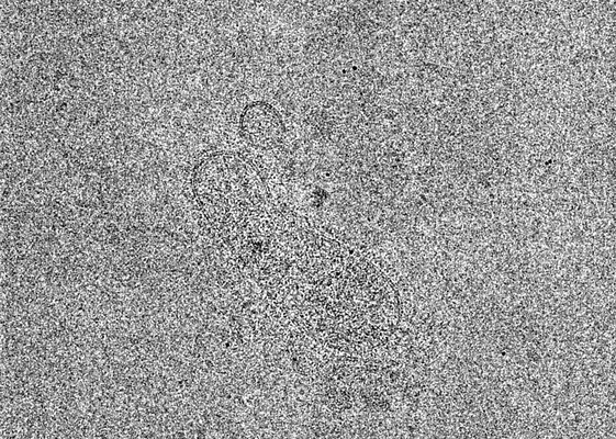 |
Cryo-ET tilt series of HPIV3 clinical isolates in the presence of an F neutralizing Fab [6 tilt series in MRC format] | Marcink T, Cheng W, Porotto M, des Georges A, Moscona A [Pubmed: 36763666] [DOI: 10.1126/sciadv.ade2727] |
19.1 GB | 16.5 Å |
| 2024-07-08 | 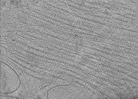 |
Structure of mavacamten-free human cardiac thick filaments within the sarcomere by cryoelectron tomography [multiple data sets in MRC format] | Chen L, Liu J, Rastegarpouyani H, Janssen PML, Pinto JR, Taylor KA [Pubmed: 38386705] [DOI: 10.1073/pnas.2311883121] |
25.6 GB | 16.5 - 26.4 Å |
| 2023-06-30 | 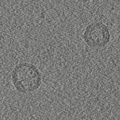 |
Cryo electron tomography of the Bacterial Voltage-Gated Sodium Channel NaChBac in Liposomes [123 tilt series in TIFF format] | Chang SYS, Kudryashev M [Pubmed: 37460541] [DOI: 10.1038/s41598-023-38027-7] |
377.3 GB | 16.3 Å |
| 2022-08-19 | 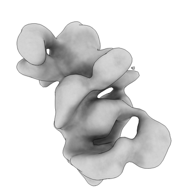 |
Single particle cryo-EM of the human CST•Polα/Primase (POLA1 FL) complex in a recruitment state [multiple data sets in MRC format] | Cai SW [Pubmed: 35578024] [DOI: 10.1038/s41594-022-00766-y] |
317.1 GB | 16.0 Å |
| 2020-04-14 | 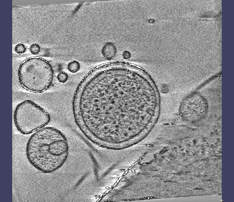 |
Cryo-electron tomography of E. coli minicells [17 multi-frame micrographs composed of 5 frames each in MRC format] | Burt A., Desfosses A., Gutsche I., Clare D. K [Pubmed: 32029744] [DOI: 10.1038/s41467-020-14350-9] |
141.7 GB | 16.0 Å |
| 2025-07-25 |  |
Cryo-electron tomography on synaptic vesicles isolated from mouse brain (part 1) [multiple data sets in TIFF and MRC formats] | Kravcenko U, Ruwolt M, Kroll J, Yushkevich A, Zenkner M, Ruta J, Lotfy R, Wanker EE, Rosenmund C, Liu F, Kudryashev M [Pubmed: 39602275] [DOI: 10.1073/pnas.2407375121] |
1.4 TB | 15.6 - 101.5 Å |
| 2020-11-20 | 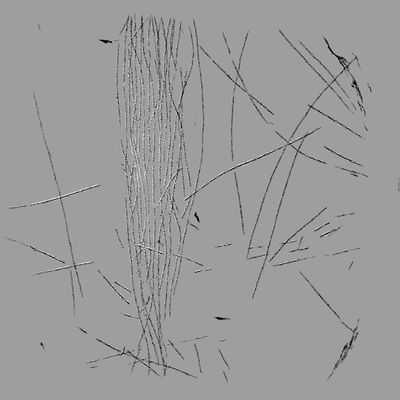 |
Cryo-ET of actin bundles induced by full length vinculin [2 tilt series in MRC format] | Ohad Medalia OM [Pubmed: 33185186] [DOI: 10.7554/eLife.53990] |
4.0 GB | 14.2 Å |
| 2021-02-19 | 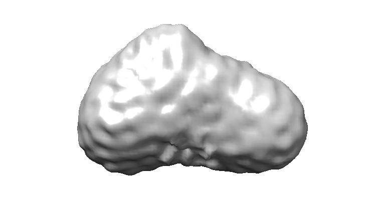 |
Negative-stained transmission electron microscopy of the nucleosome remodelling and deacetylase complex purified from murine erythroleukemia cells. [1323 micrographs in MRC format] | Mackay JP, Landsberg MJ, Jackman MJ [Pubmed: 33264611] [DOI: 10.1016/j.celrep.2020.108450] |
82.7 GB | 14.0 Å |
| 2021-02-12 | 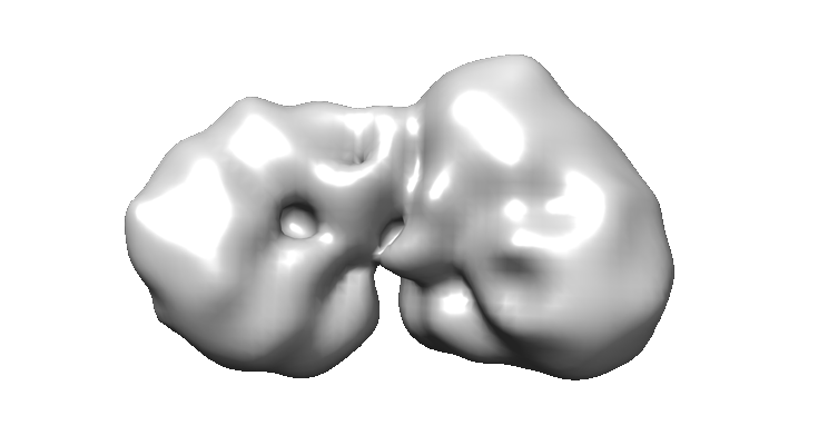 |
Negative-stained transmission electron microscopy of the nucleosome and deacetylase complex purified from murine erythroleukemia cells. [400 micrographs in MRC format] | Mackay JP, Landsberg MJ, Jackman MJ [Pubmed: 33264611] [DOI: 10.1016/j.celrep.2020.108450] |
25.0 GB | 14.0 Å |
