
Electron Microscopy Public Image Archive
Electron Microscopy Public Image Archive
Due to a storage failure, some data files are currently inaccessible (Markted: Incomplete dataset).
We are currently working to restore, but we are accepting priority requests.(email, inquiry).
We are restoring lost files from backups in the following order:
We apologize for the inconvenience and appreciate your understanding.
The EMPIAR-PDBj team at Osaka University assists Asian EM researchers with the transfer of big EM image data to EMPIAR. Instead of sending the data directly to the EBI (UK) via the internet, hard drives can also be sent to Osaka University by postal mail or via a courier service. As an alternative, internet transfer to our server in Osaka is also available. If you would like to take advantage of our submission services, please contact us first by e-mail before sending the data to us.
| Release date | Imageset | Title | Authors and references | Size | Resolution |
|---|---|---|---|---|---|
| 2021-12-23 |  |
Rabbit Muscle Aldolase 20% glycerol, 200kV [741 multi-frame micrographs composed of 96 frames each in TIFF format] | Basanta B, Hirschi M, Lander GC [Pubmed: 34981768] [DOI: 10.1107/S2059798321012110] |
282.6 GB | 3.3 Å |
| 2021-11-30 |  |
Rabbit Muscle Aldolase 20% glycerol, 300kV [2608 multi-frame micrographs composed of 50 frames each in TIFF format] | Basanta B, Hirschi M, Lander GC [Pubmed: 34981768] [DOI: 10.1107/S2059798321012110] |
580.1 GB | 4.6 Å |
| 2021-11-15 | 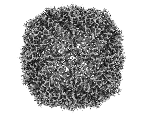 |
Apoferritin 20% glycerol, 300kV [1859 multi-frame micrographs composed of 35 frames each in TIFF format] | Basanta B, Hirschi MM, Grotjahn DA, Lander GC [Pubmed: 34981768] [DOI: 10.1107/S2059798321012110] |
477.7 GB | 2.1 Å |
| 2024-11-21 | 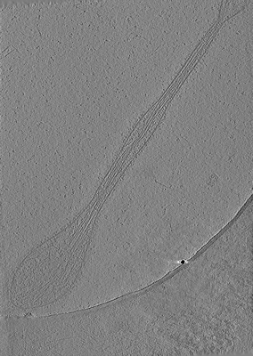 |
Human platelets in presence of SARS-CoV-2 spike protein [6 reconstructed volumes in MRC format] | Basnet N, Bodakuntla S, Nichols S, Martinez-Sanchez A, Agostini L, Soh YM, Takagi J, Biertumpfel C, Mizuno N [Pubmed: 36739444] [DOI: 10.1038/s41467-023-36279-5] |
15.3 GB | — |
| 2025-07-12 |  |
Cryo-EM structure of the wild-type Thermus thermophilus 70S ribosome in complex with mRNA, A-site Q230-N5-methylated Release Factor 1, and P-site 2'-deoxy-A76-fMEAAAKC-peptidyl-tRNAcys [multiple data sets in TIFF and MRC formats] | Basu RS, Gagnon MG, Polikanov YS [Pubmed: 40536958] [DOI: 10.1126/science.ads9030] |
4.1 TB | 2.13 Å |
| 2022-07-01 |  |
Pseudomonas aeruginosa 70S ribosome initiation complex bound to compact IF2 [8056 multi-frame micrographs composed of 31 frames each in TIFF format] | Basu RS, Sherman MB, Gagnon MG [Pubmed: 35697706] [DOI: 10.1038/s41467-022-31129-2] |
1.7 TB | 2.6 - 3.6 Å |
| 2026-03-17 |  |
Focused Ion Beam - Scanning Electron Microscopy volume of the aboral pole of a Nematostella Vectensis polyp [2347 multi-frame micrographs composed of 1 frames each in TIFF format] | Basu SH, Ronchi PR, Ikmi AI [DOI: 10.1101/2025.05.26.656114] |
16.4 GB | — |
| 2022-04-29 | 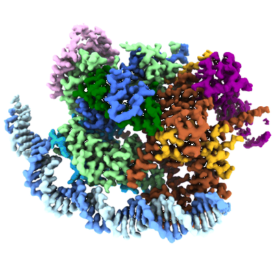 |
Structure of transcription factor UAF in complex with TBP and 35S rRNA promoter DNA [multiple data sets in TIFF format] | Baudin F, Murciano B, Fung HKH, Fromm SA, Mattei S, Mahamid J, Müller CW [Pubmed: 35442737] [DOI: 10.1126/sciadv.abn5725] |
3.1 TB | 2.8 Å |
| 2024-08-26 | 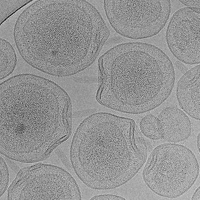 |
Molecular model of a bacterial flagellar motor in situ reveals a “parts-list” of protein adaptations to increase torque [multiple data sets in MRC format] | Beeby M, Cohen EJ, Drobnič T, Nans A, Rosenthal PB [Pubmed: 40595286] [DOI: 10.1038/s41564-025-02012-9] |
10.1 TB | 7.9 - 9.36 Å |
| 2018-02-08 |  |
Tilt-series of salmonella enterica wild-type bacterial flagellar motor [1 tilt series in MRC format] | Beeby M, Ribardo DA, Brennan CA, Ruby EG, Jensen GJ, Hendrixson DR [Pubmed: 26976588] [DOI: 10.1073/pnas.1518952113] |
328.0 MB | 69.4 Å |
| 2025-01-17 | 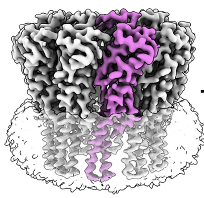 |
Cryo-EM structure of the decameric TraT surface exclusion lipoprotein from Escherichia coli (F plasmid) [8061 multi-frame micrographs composed of 40 frames each in TIFF format] | Beis K, Seddon C [DOI: 10.1101/2024.08.09.607165] |
1023.7 GB | 2.66 Å |
| 2025-01-17 |  |
Cryo-EM structure of the decameric TraT surface exclusion lipoprotein from Klebsiella pneumoniae (pKpQIL plasmid) [5558 multi-frame micrographs composed of 38 frames each in MRC format] | Beis K, Seddon C [DOI: 10.1101/2024.08.09.607165] |
6.2 TB | 2.47 Å |
| 2024-02-15 | 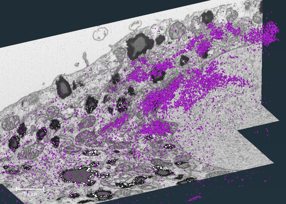 |
FIB-SEM dataset showing localization of a Golgi matrix protein GM130 in human hepatocellular carcinoma cell (Huh-7) [564 reconstructed volumes in TIFF format] | Belevich I, Jokitalo E | 1.1 GB | — |
| 2024-01-16 | 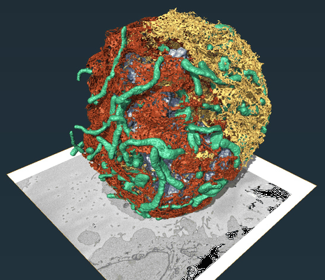 |
REEP3 and REEP4 determine the tubular morphology of the endoplasmic reticulum during mitosis [multiple data sets in DM4 and TIFF formats] | Belevich I, Jokitalo E [Pubmed: 30995177] [DOI: 10.1091/mbc.e18-11-0698] |
8.7 GB | — |
| 2024-02-15 | 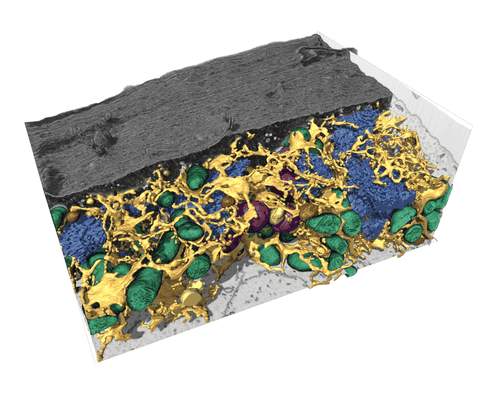 |
FIB-SEM dataset of a human bone osteosarcoma epithelial cell (U2-OS) [1168 micrographs in TIFF format] | Belevich I, Schertel A, Zaversek T, Szyrynska N, Saarnio S, Jokitalo E [Pubmed: 38609529] [DOI: 10.1038/s41556-024-01381-3] |
5.2 GB | — |
| 2026-06-01 |  |
SBF-SEM and FIB-SEM datasets and models of human macrophage and A431 cells [multiple data sets in TIFF format] | Belevich I, Szkalisity A, Vanharanta L, Ikonen E, Jokitalo E [Pubmed: 40195500] [DOI: 10.1038/s44318-025-00423-2] |
37.9 GB | — |
| 2023-01-30 | 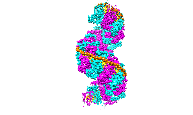 |
Structure of RecT protein from Listeria innoccua phage A118 in complex with 83-mer annealed duplex [2038 multi-frame micrographs composed of 36 frames each in MRC format] | Bell CE [Pubmed: 36543802] [DOI: 10.1038/s41467-022-35572-z] |
1.6 TB | 3.4 Å |
| 2023-01-16 | 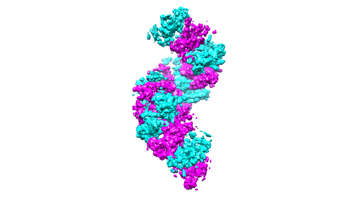 |
Structure of RecT protein from Listeria innoccua phage A118 in complex with 83-mer single stranded DNA [1619 multi-frame micrographs composed of 45 frames each in TIFF format] | Bell CE [Pubmed: 36543802] [DOI: 10.1038/s41467-022-35572-z] |
822.3 GB | 4.5 Å |
| 2022-07-12 | 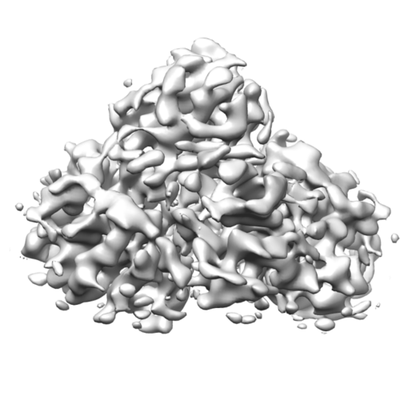 |
C2-symmetric single-particle cryo-EM map of T. vaginalis FDPF3 - unaligned multi-frame micrographs [stack of 7398 particles in TIFF format] | Bell TA [Pubmed: 35780837] [DOI: 10.1016/j.jbc.2022.102210] |
1.4 TB | 6.6 - 6.8 Å |
| 2025-10-23 |  |
3D FIB/SEM Imaging of the Molecular Layer of the Mouse Dentate Gyrus [multiple data sets in TIFF format] | Benoit LB, Pernet-Gallay KPG, Jouneau PHJ | 258.2 GB | — |
| 2021-06-11 |  |
Single particle cryo EM dataset of SARS-CoV-2 Spike protein with D614G substitution [10051 multi-frame micrographs composed of 32 frames each in MRC format] | Benton DJ, Wrobel AG, Rosenthal PB, Gamblin SJ [Pubmed: 33579792] [DOI: 10.1073/pnas.2022586118] |
4.5 TB | 3.5 - 4.2 Å |
| 2020-11-27 |  |
Micrograph frames from 110 internal SEMC/NYSBC test datasets used for Topaz-Denoise model generation & analysis [1000000 micrographs in MRCS format] | Bepler T, Kelley K, Noble AJ, Berger B [Pubmed: 33060581] [DOI: 10.1038/s41467-020-18952-1] |
33.9 TB | 2.9 - 12.54 Å |
| 2019-04-05 |  |
Rabbit muscle aldolase single particle cryoEM [multiple data sets in MRC format] | Bepler T, Morin A, Rapp M, Brasch J, Shapiro L, Noble AJ, Berger B [Pubmed: 31591578] [DOI: 10.1038/s41592-019-0575-8] |
1.4 TB | 3.7 - 3.92 Å |
| 2022-03-01 | 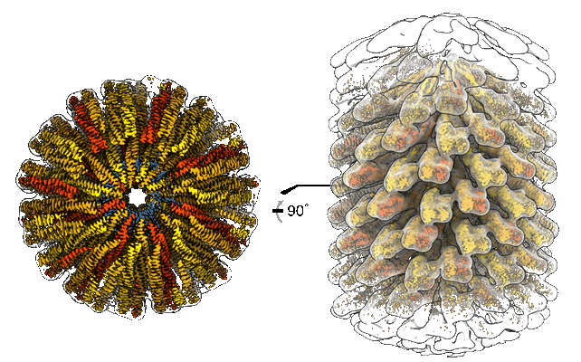 |
Helical structure of the toxin MakA from Vibrio cholera [1351 multi-frame micrographs composed of 40 frames each in TIFF format] | Berg A, Nadeem A, Uhlin BE, Wai SN, Barandun J [Pubmed: 35131030] [DOI: 10.7554/eLife.73439] |
195.8 GB | 3.65 Å |
| 2023-02-03 | 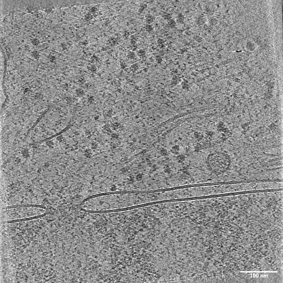 |
Cryo-electron tomography on plasma FIB lamellae of HeLa cells [6481 tilt series in EER format] | Berger C, Dumoux M, Glen T, Yee NB, Mitchels JM, Patáková Z, Darrow MC, Naismith JH, Grange M [Pubmed: 36746945] [DOI: 10.1038/s41467-023-36372-9] |
622.2 GB | 4.9 Å |
