
Electron Microscopy Public Image Archive
Electron Microscopy Public Image Archive
Due to a storage failure, some data files are currently inaccessible (Markted: Incomplete dataset).
We are currently working to restore, but we are accepting priority requests.(email, inquiry).
We are restoring lost files from backups in the following order:
We apologize for the inconvenience and appreciate your understanding.
The EMPIAR-PDBj team at Osaka University assists Asian EM researchers with the transfer of big EM image data to EMPIAR. Instead of sending the data directly to the EBI (UK) via the internet, hard drives can also be sent to Osaka University by postal mail or via a courier service. As an alternative, internet transfer to our server in Osaka is also available. If you would like to take advantage of our submission services, please contact us first by e-mail before sending the data to us.
| Release date | Imageset | Title | Authors and references | Size | Resolution |
|---|---|---|---|---|---|
| 2026-10-07 |  |
2026 CAHRA Challenge 1) Compositional Heterogeneity [5200 multi-frame micrographs composed of 945 frames each in EER format] | Feathers JR, Zhong ED, Heeter R, Woollard G, Hanson SM, Cossio P, Greer JAT, Burnley T [DOI: 10.64898/2026.09.15.751515] |
9.6 TB | — |
| 2026-10-06 |  |
2026 CAHRA Challenge 2) Conformational Heterogeneity [stack of 1753470 particles in MRCS format] | Feathers JR, Zhong ED, Heeter RC, Woollard G, Hanson SM, Cossio P, Greer JAT, Burnley T [DOI: 10.64898/2026.09.15.751515] |
215.4 GB | — |
| 2026-10-05 |  |
2026 CAHRA Challenge 3) Pose Entanglement [stack of 30000 particles in MRCS format] | Feathers JR, Zhong ED, Heeter R, Woollard G, Hanson SM, Cossio P, Greer JAT, Burnley T | 7.3 GB | — |
| 2014-01-03 | 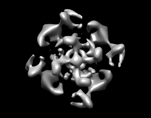 |
2D crystal images of the potassium channel MloK1 with and without cAMP ligand [multiple data sets in TIFF format] | Kowal J, Chami M, Baumgartner P, Arheit M, Chiu P-L, Rangl M, Scheuring S, Schroeder GF, Nimigean CM, Stahlberg H [Pubmed: 24469021] [DOI: 10.1038/ncomms4106] |
11.6 GB | 7.0 Å |
| 2020-07-14 | 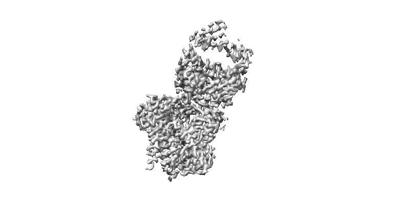 |
3 Å resolution single particle reconstruction of glucosyltransferase ALG6 in nanodisc [multiple data sets in MRC and TIFF formats] | Bloch JS, Pesciullesi G, Boilevin J, Nosol K, Irobalieva RN, Darbre T, Aebi M, Kossiakoff AA, Reymond JL, Locher KP [Pubmed: 32103179] [DOI: 10.1038/s41586-020-2044-z] |
2.8 TB | 3.0 Å |
| 2019-12-19 | 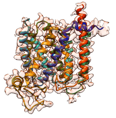 |
3.2 Å Single-Particle Cryo-EM Reconstruction of 49 kDa Membrane-Bound PfCRT Complexed with Fab [multiple data sets in MRCS and MRC formats] | Kim JK, Tan YZT, Wicht KJW, Erramilli SKE, Dhingra SKD, Okombo JO, Vendome JV, Hagenah LMH, Giacometti SIG, Warren ALW, Nosol KN, Roepe PDR, Potter CSP, Carragher BC, Kossiakoff AAK, Quick MQ, Fidock DAF, Mancia FM [Pubmed: 31776516] [DOI: 10.1038/s41586-019-1795-x] |
830.4 GB | 3.3 Å |
| 2021-03-05 | 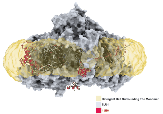 |
3.2 Å resolution structure of a functional monomeric Photosystem I from Thermosynechococcus elongatus BP-1 by single particle cryo-EM with a 200 kV CRYO ARM electron microscope [904 multi-frame micrographs composed of 60 frames each in TIFF format] | Çoruh O, Frank A, Tanaka H, Kawamoto A, El-Mohsnawy E, Kato T, Namba K, Gerle C, Nowaczyk MM, Kurisu G [Pubmed: 33686186] [DOI: 10.1038/s42003-021-01808-9] |
226.1 GB | 3.2 Å |
| 2021-05-14 | 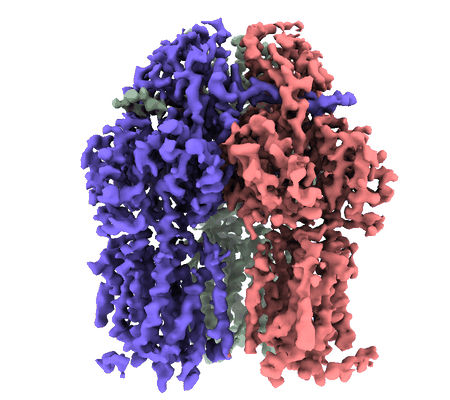 |
3.9 Angstrom reconstruction of E.coli AcrB embedded in the liposome [5757 multi-frame micrographs composed of 32 frames each in MRCS format] | Yao X, Fan X, Yan N [Pubmed: 32680969] [DOI: 10.1073/pnas.2009385117] |
2.4 TB | 3.9 Å |
| 2026-05-05 |  |
33,592 raw cryo-EM images of B. megaterium gas vesicles at 1.518 A per pixel [multiple data sets in EER, MRC and JPEG formats] | Huber ST, Jakobi AJ [Pubmed: 36868215] [DOI: 10.1016/j.cell.2023.01.041] |
16.7 TB | 3.2 Å |
| 2025-10-23 |  |
3D FIB/SEM Imaging of the Molecular Layer of the Mouse Dentate Gyrus [multiple data sets in TIFF format] | Benoit LB, Pernet-Gallay KPG, Jouneau PHJ | 258.2 GB | — |
| 2018-01-03 | 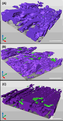 |
3D reconstruction of cardiac mitochondria in earlly prenatal (G55/57) and mid term(G58/60) and term (G66/68) GP cardiomyocyte [341 multi-frame micrographs composed of 1 frames each in TIFF format] | Kashbour H, Taggart M, White K | 1.7 GB | — |
| 2018-01-03 | 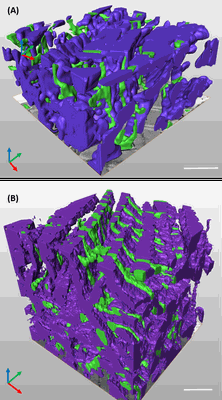 |
3D reconstruction of the cardiac mitochondria in Neaonate and Adult GP cardiomyocyte [262 multi-frame micrographs composed of 1 frames each in TIFF format] | Kashbour H, Taggart M, White K | 1.2 GB | — |
| 2018-01-03 | 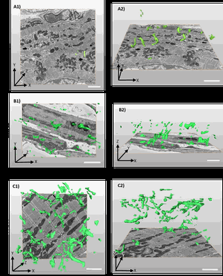 |
3D reconstruction of the tubular system of preterm G55/57, mid term G58/60 and term G66/68 of guinea pig left ventricles [318 multi-frame micrographs composed of 1 frames each in TIFF format] | Kashbour H, Taggart M | 2.9 GB | — |
| 2023-10-17 | 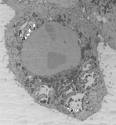 |
3D reconstructions of parasite development and the intracellular niche of the microsporidian pathogen E. intestinalis [multiple data sets in DM4 format] | Antao NVA, Lam CKL, Davydov AD, Riggi MR, Sall JS, Petzold CP, Liang FL, Iwasa JI, Ekiert DCE, Bhabha GB [Pubmed: 37425741] [DOI: 10.1101/2023.07.02.547383] |
537.9 GB | — |
| 2023-01-30 | 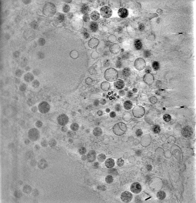 |
3D-surface reconstruction of cellular cryo-soft X-ray microscopy tomograms using semi-supervised deep learning [7 reconstructed volumes in MRC format] | Dyhr MCA, Sadeghi M, Moynova R, Knappe C, Kepsutlu Çakmak B, Werner S, Schneider G, McNally J, Noe F, Ewers H [DOI: 10.1101/2022.05.16.492055] |
23.5 GB | — |
| 2025-04-14 | 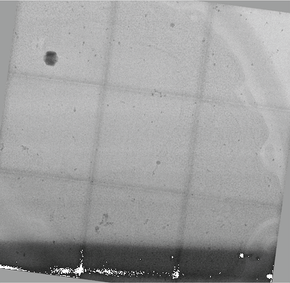 |
3x3 tiled montage tomogram of a yeast lamella imaged with a square electron beam [279 tilt series in TIFF format] | Chua EYD, Alink LM, Kopylov M, Johnston J, Eisenstein F, de Marco A [Pubmed: 38238558] [DOI: 10.1038/s41592-023-02161-x] |
58.4 GB | — |
| 2026-10-02 |  |
4 nm isotropic FIB-SEM volume of a *Planococcus citri* bacteriocyte containing nested endosymbionts, *Tremblaya princeps* and *Moranella endobia* [1 reconstructed volumes in MRC format] | Leprich DJL, Aaron JA, Hughes EH, Ladinsky MSL, Bjorkman PJB, McCutcheon JPM | 1.1 TB | — |
| 2017-08-16 | 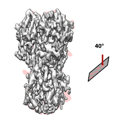 |
40 Degree Tilted Single-Particle CryoEM of Highly Preferred Orientated Influenza Hemagglutinin Trimer [multiple data sets in MRC format] | Tan YZ, Lyumkis D [Pubmed: 28671674] [DOI: 10.1038/nmeth.4347] |
354.2 GB | 4.2 Å |
| 2024-02-13 | 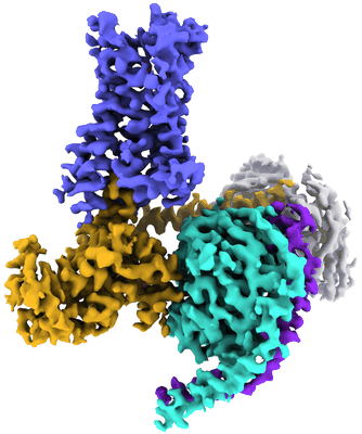 |
5-HT2AR bound to a novel agonist in complex with a mini-Gq protein and an active-state stabilizing single-chain variable fragment (scFv16) obtained by cryo-electron microscopy (cryoEM) [7573 multi-frame micrographs composed of 50 frames each in TIFF format] | Barros-Alvarez X, Kim K, Panova O, Roth BL, Skiniotis G [Pubmed: 36171289] [DOI: 10.1038/s41586-022-05258-z] |
3.4 TB | 3.45 Å |
| 2024-02-09 | 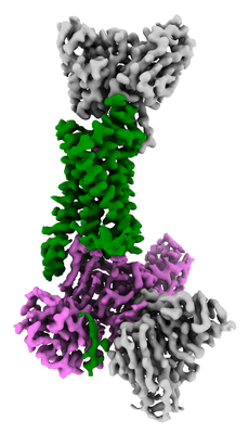 |
5-HT2B receptor bound to LSD in complex with beta-arrestin1 obtained by cryo-electron microscopy (cryoEM) [9617 multi-frame micrographs composed of 50 frames each in TIFF format] | Barros-Alvarez X [Pubmed: 36087581] [DOI: 10.1016/j.neuron.2022.08.006] |
5.0 TB | 3.3 Å |
| 2024-02-09 | 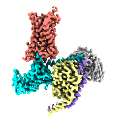 |
5-HT2B receptor bound to LSD in complex with heterotrimeric mini-Gq protein obtained by cryo-electron microscopy (cryoEM) [3916 multi-frame micrographs composed of 57 frames each in TIFF format] | Barros-Alvarez X [Pubmed: 36087581] [DOI: 10.1016/j.neuron.2022.08.006] |
2.0 TB | 2.9 Å |
| 2024-02-09 | 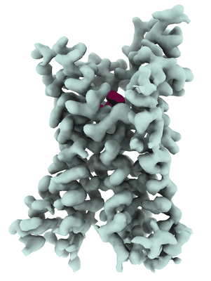 |
5-HT2B receptor bound to LSD obtained by cryo-electron microscopy (cryoEM) [12751 multi-frame micrographs composed of 50 frames each in TIFF format] | Barros-Alvarez X, Cao C, Panova O, Roth BL, Skiniotis G [Pubmed: 36087581] [DOI: 10.1016/j.neuron.2022.08.006] |
6.3 TB | 2.7 Å |
| 2025-07-30 |  |
70S initiation complex (tRNA-fMet M1 + AUG start codon) [2267 multi-frame micrographs composed of 48 frames each in TIFF format] | Mattingly JM, Nguyen HA, Roy B, Fredrick K, Dunham CM [Pubmed: 39222680] [DOI: 10.1016/j.jbc.2024.107743] |
629.3 GB | 2.75 Å |
| 2025-07-30 |  |
70S initiation complex (tRNA-fMet M1 + CUG start codon) [3155 multi-frame micrographs composed of 48 frames each in TIFF format] | Mattingly JM, Nguyen HA, Roy B, Fredrick K, Dunham CM [Pubmed: 39222680] [DOI: 10.1016/j.jbc.2024.107743] |
941.0 GB | 2.63 Å |
| 2025-07-30 |  |
70S initiation complex (tRNA-fMet M1 + GUG start codon) [2440 multi-frame micrographs composed of 48 frames each in TIFF format] | Mattingly JM, Nguyen HA, Roy B, Fredrick K, Dunham CM [Pubmed: 39222680] [DOI: 10.1016/j.jbc.2024.107743] |
705.7 GB | 2.61 Å |
