
Electron Microscopy Public Image Archive
Electron Microscopy Public Image Archive
Due to a storage failure, some datasets are currently inaccessible or incomplete (marked as Incomplete dataset).
We are pleased to report that the following entries have been restored:
EMPIAR-10577 (New)We are still accepting requests for datasets you would like us to download first (email, inquiry).
We apologize for the inconvenience and appreciate your understanding.
EMPIAR-PDBj services will be suspended during the following period for a legally required inspection of electrical equipment.
During this period, the website and data downloads from EMPIAR-PDBj will be unavailable.
Please use EMPIAR-EBI instead.
We apologize for any inconvenience.
The EMPIAR-PDBj team at Osaka University assists Asian EM researchers with the transfer of big EM image data to EMPIAR. Instead of sending the data directly to the EBI (UK) via the internet, hard drives can also be sent to Osaka University by postal mail or via a courier service. As an alternative, internet transfer to our server in Osaka is also available. If you would like to take advantage of our submission services, please contact us first by e-mail before sending the data to us.
| Release date | Imageset | Title | Authors and references | Size | Resolution |
|---|---|---|---|---|---|
| 2020-03-23 | 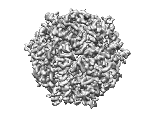 |
Micrographs of DPS collected at 100 keV using a hybrid pixel direct electron detector [739 multi-frame micrographs composed of 32 frames each in MRCS format] | Naydenova K, McMullan G, Peet MJ, Lee Y, Edwards PC, Chen S, Leahy E, Scotcher S, Henderson R, Russo CJ [Pubmed: 31709064] [DOI: 10.1107/S2052252519012612] |
23.3 GB | 3.4 Å |
| 2019-10-09 |  |
Single particle cryo-EM dataset of the triskelion hub subparticle extraction from clathrin cages [multiple data sets in MRCS format] | Morris KL, Jones JR, Halebian M, Wu S, Baker M, Armache JP, Avila Ibarra A, Sessions RB, Cameron AD, Cheng Y, Smith CJ [Pubmed: 31582853] [DOI: 10.1038/s41594-019-0292-0] |
88.4 GB | 4.69 - 7.79 Å |
| 2019-10-04 | 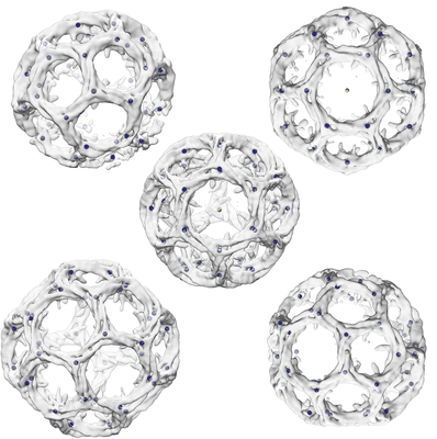 |
Single particle cryo-EM dataset of clathrin cages suitable for subparticle extraction [multiple data sets in MRCS format] | Morris KL, Jones JR, Halebian M, Wu S, Baker M, Armache JP, Avila Ibarra A, Sessions RB, Cameron AD, Cheng Y, Smith CJ [Pubmed: 31582853] [DOI: 10.1038/s41594-019-0292-0] |
19.9 GB | 9.07 - 23.68 Å |
| 2019-10-04 | 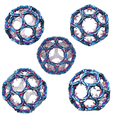 |
Single particle cryo-EM dataset of clathrin cages with phase flipping suitable for refinement [stack of 12785 particles in MRCS format] | Morris KL, Jones JR, Halebian M, Wu S, Baker M, Armache JP, Avila Ibarra A, Sessions RB, Cameron AD, Cheng Y, Smith CJ [Pubmed: 31582853] [DOI: 10.1038/s41594-019-0292-0] |
11.9 GB | 9.07 - 23.68 Å |
| 2020-02-24 | 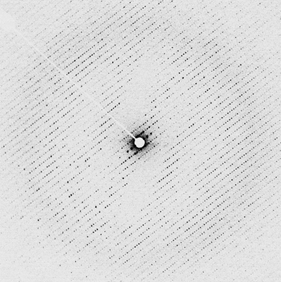 |
Electron energy-filtered diffraction (eEFD) of catalase 3D crystal with CRYO ARM 300 [84 micrographs in MRC format] | Yonekura K, Ishikawa T, Maki-Yonekura S [Pubmed: 30928615] [DOI: 10.1016/j.jsb.2019.03.009] |
5.3 GB | — |
| 2019-12-04 |  |
Cryo-EM structure of multidrug efflux pump MexAB-OprM [8722 multi-frame micrographs composed of 32 frames each in MRC format] | Tsutsumi K, Yonehara R, Ishizaka-Ikeda E, Miyazaki N, Maeda S, Iwasaki K, Nakagawa A, Yamashita E [Pubmed: 30944318] [DOI: 10.1038/s41467-019-09463-9] |
3.5 TB | 3.64 - 3.76 Å |
| 2020-02-18 | 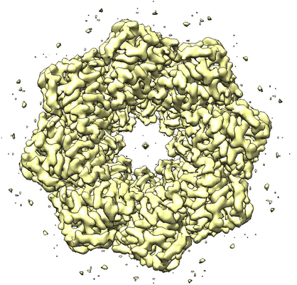 |
Structure of an undocked hemichannel of the N-terminal-deleted INX-6 in a nanodisc [300 micrographs in MRC format] | Burendei B, Shinozaki R, Watanabe M, Terada T, Tani K, Fujiyoshi Y, Oshima A [Pubmed: 32095518] [DOI: 10.1126/sciadv.aax3157] |
15.9 GB | 3.6 Å |
| 2020-02-18 | 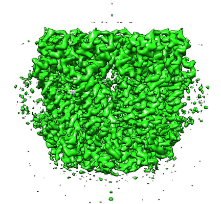 |
Cryo-EM structure of an undocked innexin-6 hemichannel in detergent [497 micrographs in MRC format] | Burendei B, Shinozaki R, Watanabe M, Terada T, Tani K, Fujiyoshi Y, Oshima A [Pubmed: 32095518] [DOI: 10.1126/sciadv.aax3157] |
26.4 GB | 3.8 Å |
| 2020-02-18 | 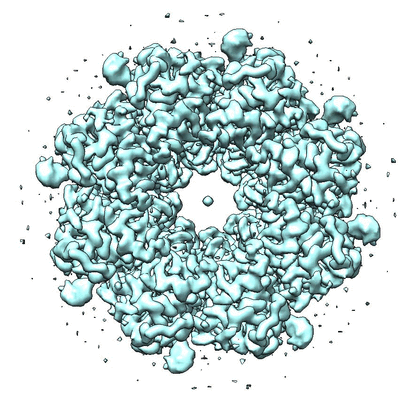 |
Cryo-EM structure of an undocked innexin-6 hemichannel in phospholipids [933 micrographs in MRC format] | Burendei B, Shinozaki R, Watanabe M, Terada T, Tani K, Fujiyoshi Y, Oshima A [Pubmed: 32095518] [DOI: 10.1126/sciadv.aax3157] |
49.5 GB | 3.8 Å |
| 2019-08-16 | 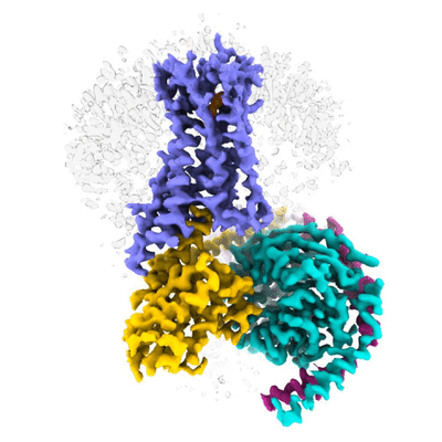 |
Cryo electron microscopy of Cannabinoid Receptor 1-G Protein Complex [2756 multi-frame micrographs composed of 40 frames each in TIFF format] | Krishna Kumar K, Shalev-Benami M, Kobilka BK, Skiniotis G [Pubmed: 30639101] [DOI: 10.1016/j.cell.2018.11.040] |
476.0 GB | 3.0 Å |
| 2020-09-11 | 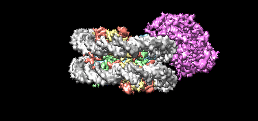 |
Cryo-EM structures of remodeler-nucleosome intermediates suggest allosteric control through the nucleosome [719 multi-frame micrographs composed of 30 frames each in MRCS format] | Armache J-P, Gamarra N, Johnson SL, Leonard JD, Wu S, Narlikar G, Cheng Y [Pubmed: 31210637] [DOI: 10.7554/eLife.46057] |
1.4 TB | 3.39 Å |
| 2019-11-19 | 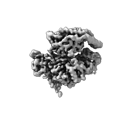 |
Cryo-electron microscopy structure of the P-Rex1–G-beta-gamma signaling scaffold [multiple data sets in TIFF, MRC and MRCS formats] | Cash JN, Urata S, Li S, Ravala SK, Avramova LV, Shost MD, Gutkind JS, Tesmer JJG, Cianfrocco MA [Pubmed: 31663027] [DOI: 10.1126/sciadv.aax8855] |
3.0 TB | 3.2 Å |
| 2019-06-19 | 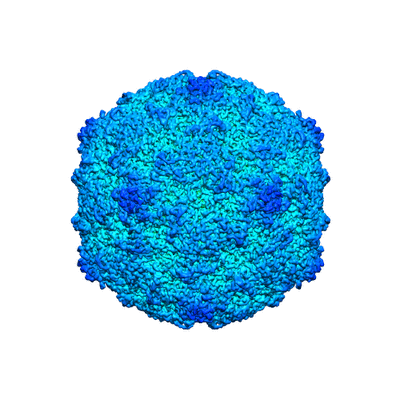 |
Extracellular albumin and endosomal ions prime enterovirus particles for uncoating that can be prevented by fatty acid saturation [multiple data sets in MRC format] | Domanska A, Ruokolainen VP, Pelliccia M, Laajala MA, Marjomäki VS, Butcher SJ [Pubmed: 31189702] [DOI: 10.1128/JVI.00599-19] |
2.4 TB | 3.5 - 3.6 Å |
| 2021-02-19 |  |
Cryo-EM structure of the mammalian ATP synthase tetramer bound to inhibitory protein IF1 (Part1) [2659 micrographs in MRC format] | Gu J, Zhang L, Zong S, Guo R, Liu T, Yi J, Wang P, Zhuo W, Yang M [Pubmed: 31197009] [DOI: 10.1126/science.aaw4852] |
141.3 GB | 3.34 - 6.2 Å |
| 2019-06-21 | 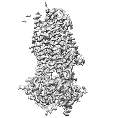 |
Cryo electron micrographs of human cystic fibrosis transmembrane conductance regulator (CFTR) in complex with GLPG [3335 multi-frame micrographs composed of 50 frames each in TIFF format] | Zhang ZZ, Liu FL, Chen JC [Pubmed: 31221859] [DOI: 10.1126/science.aaw7611] |
1.4 TB | 3.2 Å |
| 2019-11-07 |  |
Cryo-EM structure of calcium-bound TMEM16F in nanodisc with supplement of PIP2 [stack of 3823 particles in MRCS format] | Feng S, Jan LY, Cheng Y [Pubmed: 31291589] [DOI: 10.1016/j.celrep.2019.06.023] |
576.6 GB | 3.2 - 3.3 Å |
| 2020-04-21 | 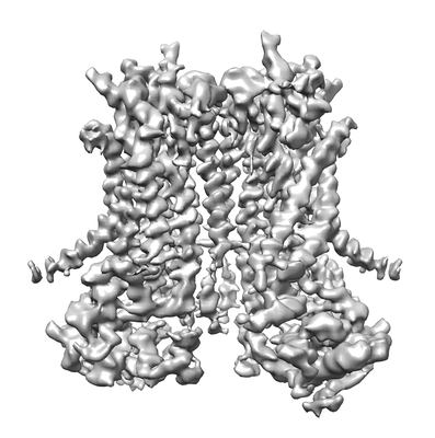 |
Cryo-EM structure of TMEM16F in digitonin without calcium bound [stack of 2249 particles in MRCS format] | Feng S, Dang S, Han T, Ye W, Jin P, Cheng T, Li J, Jan YN, Jan LY, Cheng Y [Pubmed: 31291589] [DOI: 10.1016/j.celrep.2019.06.023] |
289.5 GB | 3.9 Å |
| 2020-06-30 | 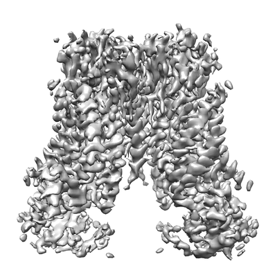 |
Cryo-EM structure of TMEM16F in digitonin with calcium bound [stack of 2505 particles in MRC format] | Feng S, Dang S, Han TW, Ye W, Jin P, Cheng T, Li J, Jan YN, Jan LY, Cheng Y [Pubmed: 31291589] [DOI: 10.1016/j.celrep.2019.06.023] |
308.8 GB | 3.5 Å |
| 2020-03-02 | 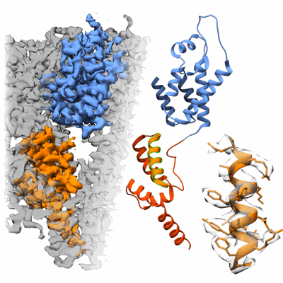 |
Tilt-schemes benchmarking for cryo electron tomography of HIV-1 CA-SP1 [multiple data sets in MRC format] | Turoňová B, Hagen WJH, Obr M, Mosalaganti S, Beugelink JW, Zimmerli CE, Kräusslich HG, Beck M [Pubmed: 32054835] [DOI: 10.1038/s41467-020-14535-2] |
45.6 GB | 4.2 Å |
| 2019-12-19 |  |
Structure of a synthetic beta-carboxysome shell [1343 multi-frame micrographs composed of 25 frames each in TIFF format] | Laughlin TG [Pubmed: 31501298] [DOI: 10.1104/pp.19.00885] |
423.5 GB | 2.6 - 4.1 Å |
| 2019-07-29 | 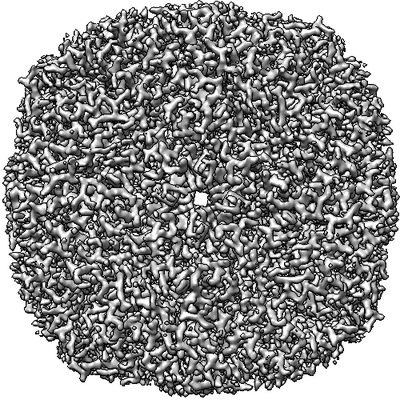 |
Movies of horse spleen apoferritin on multifunctional ultrastable graphene supports for electron cryomicroscopy [480 multi-frame micrographs composed of 38 frames each in MRCS format] | Naydenova K, Peet MJ, Russo CJ [Pubmed: 31127045] [DOI: 10.1073/pnas.1904766116] |
570.0 GB | 2.1 Å |
| 2019-10-17 |  |
Cryo electron microscopy of TssA protein from T6SS of Vibrio cholerae. [multiple data sets in MRC and MRCS formats] | Nazarov S, Adaixo R, Basler M [Pubmed: 31403721] [DOI: 10.15252/embj.2018100825] |
1.3 TB | 3.9 Å |
| 2019-06-07 | 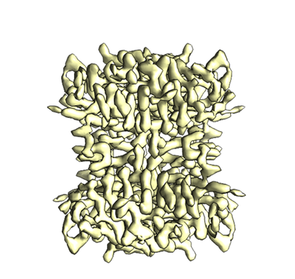 |
Single particle reconstruction of 52 kDa biotin-bound state streptavidin at 3.2 Angstrom resolution [3309 multi-frame micrographs composed of 32 frames each in MRC format] | Fan X, Wang J, Zhang X, Yang Z, Zhang JC, Zhao L, Peng HL, Lei J, Wang HW [Pubmed: 31160591] [DOI: 10.1038/s41467-019-10368-w] |
5.5 TB | 3.2 Å |
| 2019-06-07 |  |
Single particle reconstruction of 52 kDa apo-state streptavidin at 3.3 Angstrom resolution [1448 multi-frame micrographs composed of 32 frames each in MRC format] | Fan X, Wang J, Zhang X, Yang Z, Zhang JC, Zhao L, Peng HL, Lei J, Wang HW [Pubmed: 31160591] [DOI: 10.1038/s41467-019-10368-w] |
2.4 TB | 3.3 Å |
| 2020-07-14 | 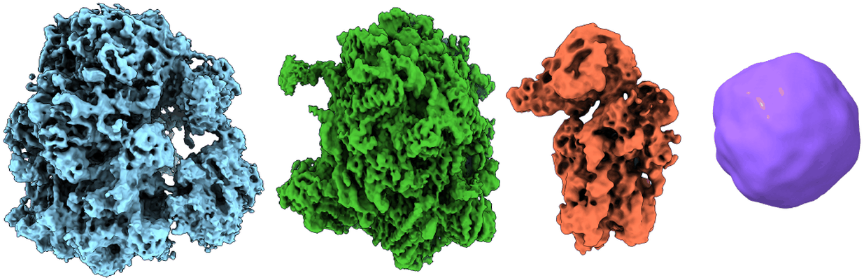 |
Separating distinct macromolecular assemblies from cryo-EM images [2423 micrographs in MRC format] | Verbeke EJ, Zhou Y, Horton AP, Mallam AL, Taylor DW, Marcotte EM [Pubmed: 31726096] [DOI: 10.1016/j.jsb.2019.107416] |
128.5 GB | 4.0 - 19.0 Å |
