
Electron Microscopy Public Image Archive
Electron Microscopy Public Image Archive
Due to a storage failure, some data files are currently inaccessible (Markted: Incomplete dataset).
We are currently working to restore, but we are accepting priority requests.(email, inquiry).
We are restoring lost files from backups in the following order:
We apologize for the inconvenience and appreciate your understanding.
The EMPIAR-PDBj team at Osaka University assists Asian EM researchers with the transfer of big EM image data to EMPIAR. Instead of sending the data directly to the EBI (UK) via the internet, hard drives can also be sent to Osaka University by postal mail or via a courier service. As an alternative, internet transfer to our server in Osaka is also available. If you would like to take advantage of our submission services, please contact us first by e-mail before sending the data to us.
| Release date | Imageset | Title | Authors and references | Size | Resolution |
|---|---|---|---|---|---|
| 2026-05-24 |  |
Two-dimensional analysis of rat CRP [21 micrographs in MRC format] | Cheng B, Lv JM, Liang YL, Zhu L, Huang XP, Li HY, Potempa LA, Ji SR, Wu Y [Pubmed: 35338247] [DOI: 10.1038/s42003-022-03220-3] |
1.3 GB | — |
| 2022-12-13 | 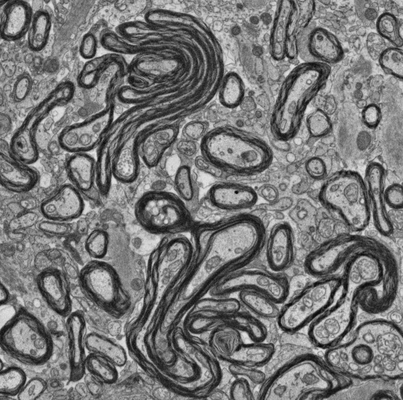 |
Focused ion beam-scanning electron microscopy links pathological myelin outfoldings to axonal changes in mice lacking Plp1 or Mag [1151 micrographs in TIFF format] | Steyer AM, Möbius W [Pubmed: 36354016] [DOI: 10.1002/glia.24290] |
17.8 GB | — |
| 2024-11-21 |  |
Interleaved scanning electron microscopy: exploring native samples at the mesoscale. E.gracilis [multiple data sets in TIFF and MRC formats] | Dumoux M | 249.7 GB | — |
| 2020-12-11 | 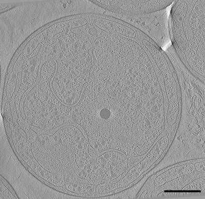 |
Cryo electron tomography after FIB-milling of Planctomycetes species Tuwongella immobilis [121 tilt series in MRC format] | Seeger C, Andersson SG [Pubmed: 32761170] [DOI: 10.1093/gbe/evaa159] |
3.2 GB | — |
| 2026-05-24 |  |
Two-dimensional analysis of mouse CRP [22 micrographs in MRC format] | Cheng B, Lv JM, Liang YL, Zhu L, Huang XP, Li HY, Potempa LA, Ji SR, Wu Y [Pubmed: 35338247] [DOI: 10.1038/s42003-022-03220-3] |
1.3 GB | — |
| 2022-12-13 | 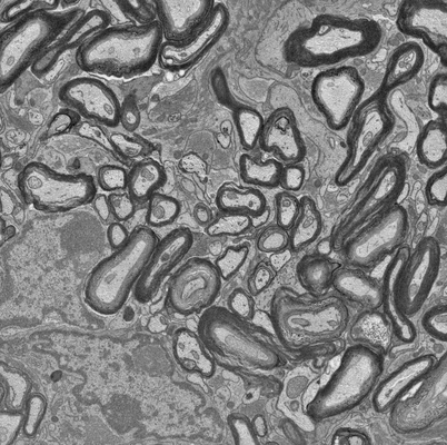 |
Focused ion beam-scanning electron microscopy links pathological myelin outfoldings to axonal changes in mice lacking Plp1 or Mag [1447 micrographs in TIFF format] | Steyer AM, Möbius W [Pubmed: 36354016] [DOI: 10.1002/glia.24290] |
34.6 GB | — |
| 2025-02-04 | 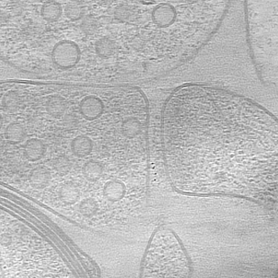 |
Cryo-electron tomographic investigation of native hippocampal glutamatergic synapses [multiple data sets in TIFF format] | Matsui A, Spangler CJ, Elferich J, Gouaux E [Pubmed: 39495821] [DOI: 10.7554/eLife.98458] |
9.5 GB | — |
| 2020-12-11 | 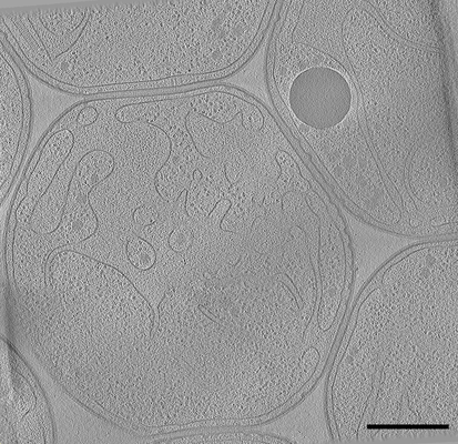 |
Cryo electron tomography after FIB-milling of Planctomycetes species Gemmata obscuriglobus (#1) [101 tilt series in MRC format] | Seeger C, Andersson SG [Pubmed: 32761170] [DOI: 10.1093/gbe/evaa159] |
2.7 GB | — |
| 2020-12-11 | 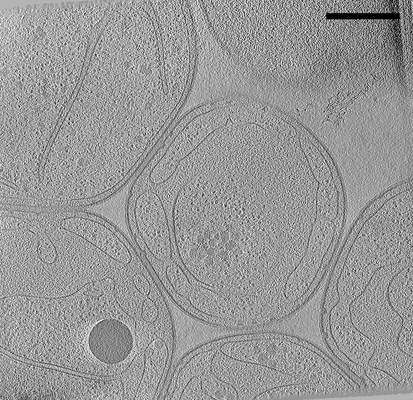 |
Cryo electron tomography after FIB-milling of Planctomycetes species Gemmata obscuriglobus (#2) [101 tilt series in MRC format] | Seeger C, Andersson SG [Pubmed: 32761170] [DOI: 10.1093/gbe/evaa159] |
2.7 GB | — |
| 2025-02-04 |  |
Microtubule plus end architecture in presence of the ciliary tip module. [34 tilt series in TIFF format] | Saunders H.A.J., Van den Berg C.M., Hoogebeen R.A., Schweizer D., Stecker K.E., Roepman R., Howes S.C., Akhmanova A. | 58.0 GB | — |
| 2026-02-24 |  |
CryoEM data of alpha-synuclein fibril with tetrazine 2.0 at site 60 [7815 micrographs in MRC format] | Jenkins RA, Rodriguez JA, Sawaya MR | 250.8 GB | — |
| 2018-07-05 | 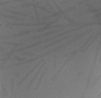 |
Cryo-EM structure of alpha-synuclein fibrils [118 multi-frame micrographs composed of 50 frames each in MRC format] | Guerrero-Ferreira R, Taylor NM, Mona D, Ringler P, Lauer ME, Riek R, Britschgi M, Stahlberg H [Pubmed: 29969391] [DOI: 10.7554/elife.36402] |
6.3 GB | — |
| 2020-12-11 | 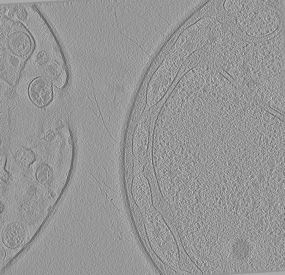 |
Cryo electron tomography after FIB-milling of Planctomycetes species Tuwongella immobilis (33k magnification) [multiple data sets in MRC format] | Seeger C, Andersson SG [Pubmed: 32761170] [DOI: 10.1093/gbe/evaa159] |
15.5 GB | — |
| 2022-12-13 | 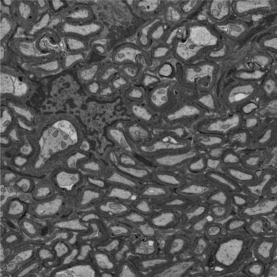 |
Focused ion beam-scanning electron microscopy links pathological myelin outfoldings to axonal changes in mice lacking Plp1 or Mag [940 micrographs in TIFF format] | Steyer AM, Möbius W [Pubmed: 36354016] [DOI: 10.1002/glia.24290] |
12.5 GB | — |
| 2025-04-07 |  |
Dynamic microtubule plus end architecture in presence of EB3. [51 tilt series in TIFF format] | Saunders H.A.J., Van den Berg C.M., Hoogebeen R.A., Schweizer D., Stecker K.E., Roepman R., Howes S.C., Akhmanova A. [Pubmed: 39856351] [DOI: 10.1038/s41594-025-01483-y] |
75.9 GB | — |
| 2022-12-13 | 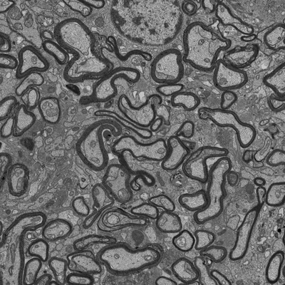 |
Focused ion beam-scanning electron microscopy links pathological myelin outfoldings to axonal changes in mice lacking Plp1 or Mag [1290 micrographs in TIFF format] | Steyer AM, Möbius W [Pubmed: 36354016] [DOI: 10.1002/glia.24290] |
22.4 GB | — |
| 2024-12-02 |  |
Interleaved scanning electron microscopy: exploring native samples at the mesoscale.Adult mouse brain [multiple data sets in TIFF and MRC formats] | Dumoux M | 344.8 GB | — |
| 2018-07-06 | 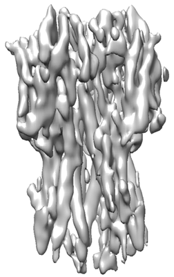 |
Single particle cryoEM of hemagglutinin with spot-to-plunge time of 500ms [multiple data sets in MRC format] | Noble AJ, Wei H, Dandey VP, Zhang Z, Potter CS, Carragher B [Pubmed: 30250056] [DOI: 10.1038/s41592-018-0139-3] |
51.1 GB | — |
| 2021-05-21 | 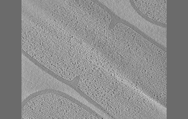 |
Tilt series of dividing vegetative and sporulating cells of Bacillus subtilis from the manuscript - Khanna et al., 2021 [multiple data sets in MRC format] | Khanna K, Lopez-Garrido J, Sugie J, Pogliano K, Villa E [Pubmed: 34018921] [DOI: 10.7554/eLife.62204] |
8.7 GB | — |
| 2025-12-17 |  |
Raw data for cryo-tomograms of Agrobacterium phage 7-7-1 and Agrobacterium sp. H13-3 [841 tilt series in MRC format] | Noteborn WEM, Hoeksma T, Rouchen OY, Briegel A [Pubmed: 41353512] [DOI: 10.1038/s42003-025-09319-7] |
194.7 GB | — |
| 2026-04-20 |  |
Cryo electron tomography data presented in Multi-scale Molecular Imaging of Human Cells reveals COPI and COPII Vesicles at ER Exit Sites [multiple data sets in MRC format] | Downes K, Zanetti G [DOI: 10.1101/2025.07.29.667472] |
637.7 GB | — |
| 2022-03-14 | 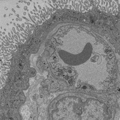 |
SBF SEM of Human term placental villi [multiple data sets in TIFF format] | Lewis RM [DOI: 10.1101/2022.01.26.477815] |
90.2 GB | — |
| 2025-10-23 |  |
Raw micrographs of apoferritin specimen sprayed onto precooled grid coated in liquid ethane [466 multi-frame micrographs composed of 1998 frames each in EER format] | Gusach A, Russo CJ, Sader K [DOI: 10.1073/pnas.2516900122] |
332.0 GB | — |
| 2026-05-19 |  |
Aquifex aeolicus lumazine synthase in complex with riboflavin synthase [8625 multi-frame micrographs composed of 40 frames each in TIFF format] | Koziej L, Azuma Y [DOI: 10.1038/s41467-026-73260-4] |
3.1 TB | — |
| 2026-06-19 |  |
Rabbit Aldolase, Laser Phase Plate TEM, Laser-ON dataset 1 (A1) [79 multi-frame micrographs composed of 16384 frames each in EER format] | Petrov P.N., Zhang J.T., Remis J.P., Axelrod J.A., Chen HC, Hicklin I.K., Sandhaus S, Schnurr C, Glaeser R.M., Mueller H.M. [Pubmed: 42275466] [DOI: 10.1126/science.aeh0665] |
69.7 GB | — |
