
Electron Microscopy Public Image Archive
Electron Microscopy Public Image Archive
Due to a storage failure, some datasets are currently inaccessible or incomplete (marked as Incomplete dataset).
We are pleased to report that the following entries have been restored:
EMPIAR-10577 (New)We are still accepting requests for datasets you would like us to download first (email, inquiry).
We apologize for the inconvenience and appreciate your understanding.
EMPIAR-PDBj services will be suspended during the following period for a legally required inspection of electrical equipment.
During this period, the website and data downloads from EMPIAR-PDBj will be unavailable.
Please use EMPIAR-EBI instead.
We apologize for any inconvenience.
The EMPIAR-PDBj team at Osaka University assists Asian EM researchers with the transfer of big EM image data to EMPIAR. Instead of sending the data directly to the EBI (UK) via the internet, hard drives can also be sent to Osaka University by postal mail or via a courier service. As an alternative, internet transfer to our server in Osaka is also available. If you would like to take advantage of our submission services, please contact us first by e-mail before sending the data to us.
| Release date | Imageset | Title | Authors and references | Size | Resolution |
|---|---|---|---|---|---|
| 2016-09-21 | 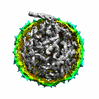 |
Asymmetric cryo-EM reconstruction of phage MS2 reveals genome structure in situ [751 micrographs in MRC format] | Koning RI, Gomez-Blanco J, Akopjana I, Vargas J, Kazaks A, Tars K, Carazo JM, Koster AJ [Pubmed: 27561669] [DOI: 10.1038/ncomms12524] |
46.9 GB | 5.0 - 10.5 Å |
| 2022-01-31 | 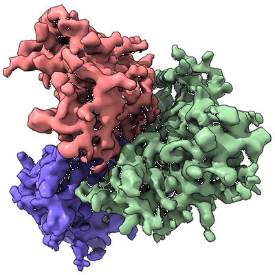 |
Asymmetric structures of the uncleaved full-length HIV-1 envelope glycoprotein trimer [multiple data sets in MRC and BIG DATA VIEWER HDF5 formats] | Zhang S, Wang KY, Wang WL, Chen S, Sodroski JG, Mao Y [Pubmed: 34549974] [DOI: 10.1128/JVI.00529-21] |
4.0 TB | 4.1 - 4.7 Å |
| 2020-05-27 | 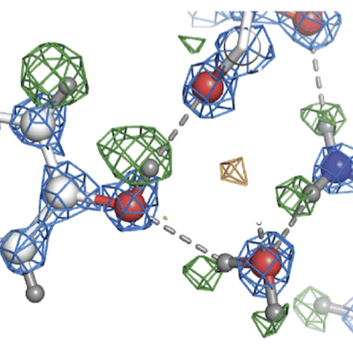 |
Atomic resolution structure of apoferritin [3370 multi-frame micrographs composed of 434 frames each in EER format] | Nakane T, Kotecha A, Sente A, McMullan G, Masiulis S, Brown PMGE, Grigoras IT, Malinauskaite L, Malinauskas T, Miehling J, Uchański T, Yu L, Karia D, Pechnikova EV, de Jong E, Keizer J, Bischoff M, McCormack J, Tiemeijer P, Hardwick SW, Chirgadze DY, Murshudov G, Aricescu AR, Scheres SHW [Pubmed: 33087931] [DOI: 10.1038/s41586-020-2829-0] |
538.1 GB | 1.22 Å |
| 2021-02-12 | 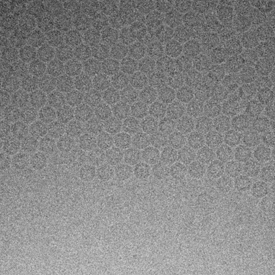 |
Atomic resolution structure of apoferritin from Titan Mono/BCorr microscope [43292 multi-frame micrographs composed of 40 frames each in MRC format] | Yip KM, Fischer N, Paknia E, Chari A, Stark H [Pubmed: 33087927] [DOI: 10.1038/s41586-020-2833-4] |
18.7 TB | 1.15 - 1.56 Å |
| 2024-06-13 | 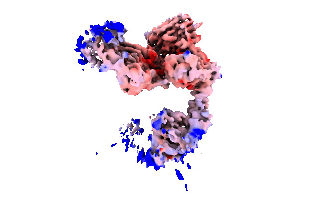 |
Atomic structure and conformational variability of the HER2-trastuzumab-pertuzumab complex [stack of 708609 particles in MRCS format] | Ruedas R, Vuillemot R, Tubiana T, Winter JM, Pieri L, Arteni AA, Samson C, Jonic S, Mathieu M, Bressanelli S [Pubmed: 38723875] [DOI: 10.1016/j.jsb.2024.108095] |
207.2 GB | 3.01 - 3.33 Å |
| 2022-04-19 | 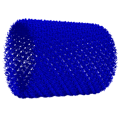 |
Atomic structure of Lanreotide nanotubes revealed by cryo-EM [stack of 631121 particles in MRCS format] | Pieri L, Wang F, Bressanelli S, Egelman EH, Paternostre M [Pubmed: 35042822] [DOI: 10.1073/pnas.2120346119] |
362.5 GB | 2.46 Å |
| 2023-10-06 | 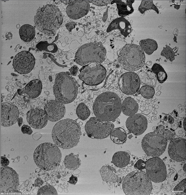 |
Atypical flagella assembly and haploid genome coiling during male gamete formation in Plasmodium [multiple data sets in MRC format] | Hair M [DOI: 10.1101/2023.05.17.540968] |
100.7 GB | — |
| 2024-07-03 |  |
Automatically collected tilt series on cryoFIB milled lamellae of Baker's Yeast [multiple data sets in TIFF and MRC formats] | Eisenstein F, Fukuda Y, Danev R [Pubmed: 39117874] [DOI: 10.1038/s41592-024-02373-9] |
1.2 TB | — |
| 2024-11-08 | 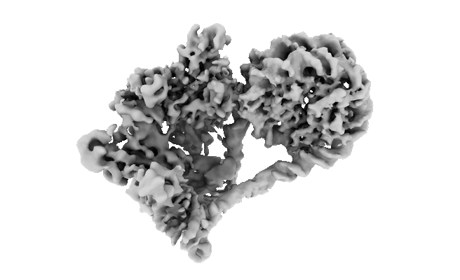 |
Automethylated PRC2 dimer bound to nucleosome [multiple data sets in MRC format] | Sauer PV, Pavlenko E, Nogales E, Poepsel S, Cookis T [Pubmed: 39303719] [DOI: 10.1016/j.molcel.2024.08.025] |
1.8 TB | 4.0 - 6.2 Å |
| 2026-04-20 |  |
BG505.SOSIP.664.T332N gp140 trimer in complex with 44m bnAb [4916 multi-frame micrographs composed of 20 frames each in MRC format] | Dutta S [Pubmed: 37649696] [DOI: 10.1016/j.isci.2023.107579] |
733.3 GB | 4.4 Å |
| 2025-07-25 |  |
BSEP Apo Structure in GDN [17482 multi-frame micrographs composed of 33 frames each in TIFF format] | Reddy BG, Gruget C, Moore JM [Pubmed: 40195555] [DOI: 10.1038/s42003-025-07908-0] |
1.4 TB | 2.8 Å |
| 2025-07-25 |  |
BSEP E297G Structure in GDN [12223 multi-frame micrographs composed of 36 frames each in TIFF format] | Reddy BG, Gruget C, Moore JM [Pubmed: 40195555] [DOI: 10.1038/s42003-025-07908-0] |
1.7 TB | 3.23 Å |
| 2024-06-17 | 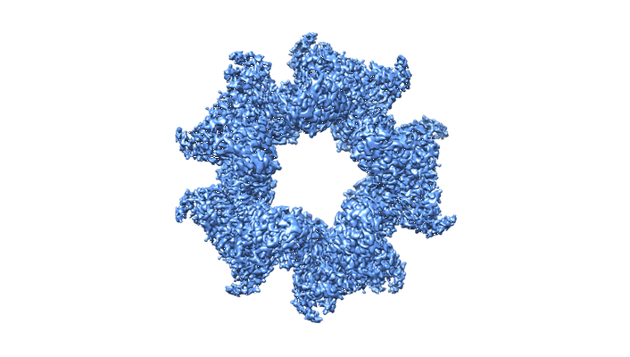 |
Bacterial selenocysteine synthase structure revealed by single-particle cryoEM [stack of 4592 particles in TIFF format] | Balasco Serrao VH, Minari K, Pereira HM, Thiemann OH [Pubmed: 38681238] [DOI: 10.1016/j.crstbi.2024.100143] |
24.6 TB | 2.69 Å |
| 2017-03-13 | 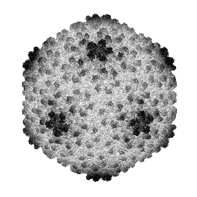 |
Bacteriophage P22 mature virion capsid protein [stack of 45150 particles in IMAGIC format] | Hryc CF, Chen D-H, Afonine PV, Jakana J, Wang Z, Haase-Pettingell C, Jiang W, Adams PD, King JA, Schmid MF, Chiu W [Pubmed: 28270620] [DOI: 10.1073/pnas.1621152114] |
159.4 GB | 3.3 Å |
| 2021-06-18 | 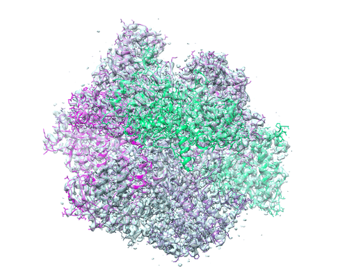 |
Bacteriophage PhiKZ non-virion RNA Polymerase [10741 multi-frame micrographs composed of 32 frames each in TIFF format] | de Martin Garrido N, Lai Wan Loong YTE, Aylett CHS [Pubmed: 34181731] [DOI: 10.1093/nar/gkab539] |
3.3 TB | 3.3 Å |
| 2021-06-04 | 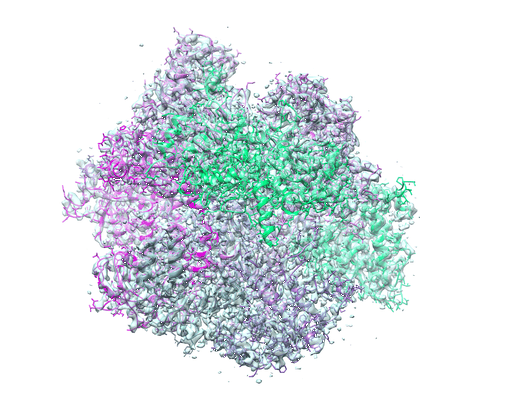 |
Bacteriophage PhiKZ non-virion RNA Polymerase [8765 micrographs in MRC format] | de Martin Garrido N, Lai Wan Loong YTE, Aylett CHS [Pubmed: 34181731] [DOI: 10.1093/nar/gkab539] |
769.6 GB | 3.3 Å |
| 2019-02-01 | 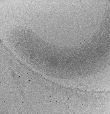 |
Bdellovibrio bacteriovorus electron cryotomography tilt-series acquired by fast-incremental method [1 tilt series in MRC format] | Chreifi G, Chen S, Metskas LA, Kaplan M, Jensen GJ [Pubmed: 30639925] [DOI: 10.1016/j.jsb.2018.12.008] |
5.2 GB | — |
| 2019-02-01 | 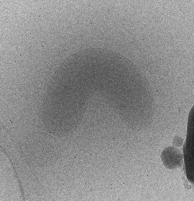 |
Bdellovibrio electron cryotomography tilt-series acquired by continuous tilting [1 tilt series in MRC format] | Chreifi G, Chen S, Metskas LA, Kaplan M, Jensen GJ [Pubmed: 30639925] [DOI: 10.1016/j.jsb.2018.12.008] |
2.1 GB | — |
| 2023-05-22 | 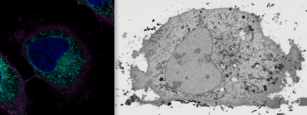 |
Benchmark FIB SEM data (#2) of HeLa cells previously imaged by Zeiss LSM900 Airyscan microscopy [multiple data sets in TIFF format] | Peddie CJ, Domart MC, Collinson LM [DOI: 10.1101/2023.05.11.540445] |
511.9 GB | — |
| 2021-10-29 | 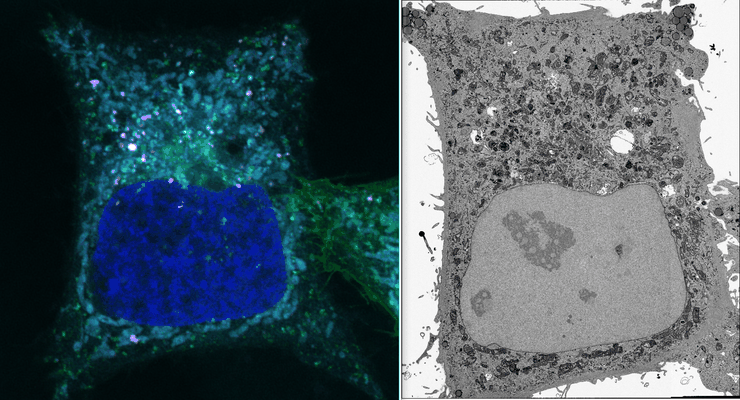 |
Benchmark FIB SEM data of HeLa cells previously imaged by Zeiss LSM900 Airyscan microscopy [multiple data sets in TIFF format] | Peddie CJ, Domart MC, Collinson L | 1.7 TB | — |
| 2023-09-05 | 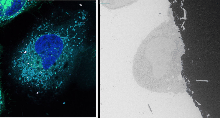 |
Benchmark SBF SEM data of HeLa cells previously imaged by Zeiss LSM900 Airyscan microscopy [multiple data sets in DM4 and TIFF formats] | Domart MC, Collinson LM [DOI: 10.1101/2023.05.11.540445] |
39.2 GB | — |
| 2025-09-05 |  |
Benchmark TEM data of mouse brain previously imaged by Nikon confocal microscopy [1 micrographs in TIFF format] | Konishi KK, Neves GN, Russell MR, Burrone JB, Fleck RF [DOI: 10.1111/jmi.13436] |
2.5 GB | — |
| 2025-08-29 |  |
Benchmarking Talos Arctica with K3 and Biocontinuum energy filter [1483 multi-frame micrographs composed of 50 frames each in TIFF format] | Maruthi K, Morais M | 334.9 GB | 1.88 Å |
| 2018-06-13 | 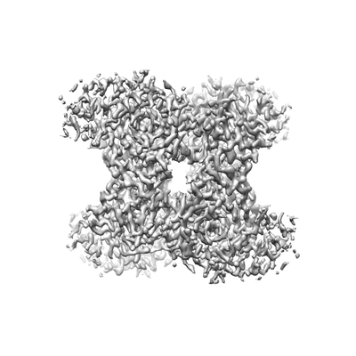 |
Benchmarking cryo-EM single particle analysis workflow [1614 multi-frame micrographs composed of 30 frames each in MRC format] | Kim LY, Rice WJ, Eng ET, Kopylov M, Cheng A, Raczkowski AM, Jordan KD, Bobe D, Potter CS, Carragher B [Pubmed: 29951483] [DOI: 10.3389/fmolb.2018.00050] |
85.6 GB | 2.4 - 2.8 Å |
| 2018-05-11 | 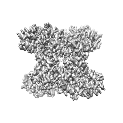 |
Benchmarking cryo-EM single particle analysis workflow [1626 multi-frame micrographs composed of 30 frames each in MRC format] | Kim LK, Rice WJ, Eng ET, Kopylov M, Cheng A, Raczkowski AM, Jordan KJ, Bobe D, Potter CS, Carragher B [Pubmed: 29951483] [DOI: 10.3389/fmolb.2018.00050] |
345.0 GB | 3.0 - 4.6 Å |
