
Electron Microscopy Public Image Archive
Electron Microscopy Public Image Archive
Due to a storage failure, some data files are currently inaccessible (Markted: Incomplete dataset).
We are currently working to restore, but we are accepting priority requests.(email, inquiry).
We are restoring lost files from backups in the following order:
We apologize for the inconvenience and appreciate your understanding.
The EMPIAR-PDBj team at Osaka University assists Asian EM researchers with the transfer of big EM image data to EMPIAR. Instead of sending the data directly to the EBI (UK) via the internet, hard drives can also be sent to Osaka University by postal mail or via a courier service. As an alternative, internet transfer to our server in Osaka is also available. If you would like to take advantage of our submission services, please contact us first by e-mail before sending the data to us.
| Release date | Imageset | Title | Authors and references | Size | Resolution |
|---|---|---|---|---|---|
| 2021-02-12 | 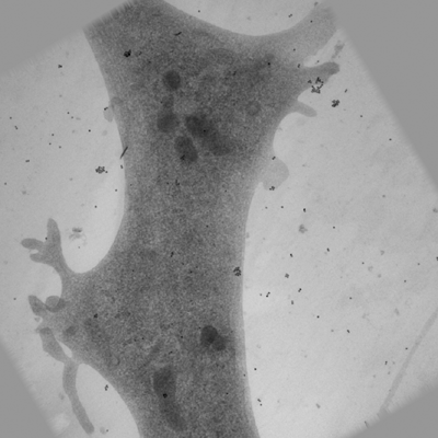 |
Ultra-high voltage electron microscope tomography tilt series of 0.7-μm-thick neurite section acquired at 15,000× magnification at an accelerating voltage of 1 MV [1 tilt series in MRC format] | Nishida T, Yoshimura R, Nishi R, Imoto Y, Endo Y [Pubmed: 32133640] [DOI: 10.1111/jmi.12885] |
120.3 MB | — |
| 2025-07-08 |  |
FIB-SEM Nicotiana benthamiana epidermal cell [multiple data sets in TIFF format] | Czymmek KJ, Burch-Smith T, Hurník Konečná T [DOI: 10.1016/bs.mcb.2023.04.008] |
623.5 GB | — |
| 2025-12-17 |  |
Cryo ET of Native Hippocampal Glutamatergic Synapses Using Dimeric AuNP Labeling [multiple data sets in EER format] | Kim H, Spangler CJ, Matsui A, Elferich J, Gouaux E, Kim J, Roseborough A, Nyman M [Pubmed: 41284882] [DOI: 10.1073/pnas.2524034122] |
18.4 GB | — |
| 2017-11-28 | 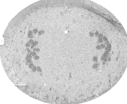 |
FIB-SEM of a dividing cell at 3.1 min after anaphase onset [1652 multi-frame micrographs composed of 1 frames each in TIFF format] | Otsuka S, Steyer AM, Schorb M, Hériché JK, Hossain MJ, Sethi S, Kueblbeck M, Schwab Y, Beck M, Ellenberg J [Pubmed: 29323269] [DOI: 10.1038/s41594-017-0001-9] |
27.0 GB | — |
| 2020-05-27 | 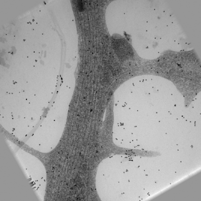 |
Ultra-high voltage electron microscope tomography tilt series of 0.7-μm-thick neurite section acquired at 20,000× magnification at an accelerating voltage of 1 MV [1 tilt series in MRC format] | Nishida T, Yoshimura R, Nishi R, Imoto Y, Endo Y [Pubmed: 32133640] [DOI: 10.1111/jmi.12885] |
120.3 MB | — |
| 2023-02-22 | 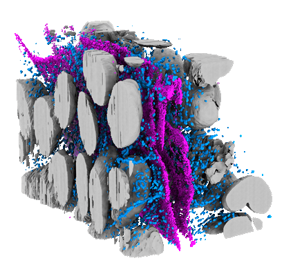 |
Light and electron microscopy continuum-resolution imaging of 3D cell cultures [19 multi-frame micrographs composed of 1 frames each in MRC format] | DImprima EDI, Garcia Montero MGM, Gawrzak, SG, Ronchi PR, Zagoriy IZ, Schwab YS, Jechlinger JS, Mahamid JM | 72.9 GB | — |
| 2025-06-30 |  |
FIB-SEM Chinese Hamster Ovary cell line sample dataset [multiple data sets in TIFF and PNG formats] | Slamková D., Hurník Konečná T [DOI: 10.1016/bs.mcb.2023.04.008] |
1.3 GB | — |
| 2025-12-24 |  |
Cryogenic transmission electron microscopy images of extracellular vesicles isolated from conditioned media of pancreatic cancer and normal pancreatic epithelial cell lines [3 micrographs in TIFF format] | Singh BS, Gaur PG, Bose PB, Zhang YJ, Li YL, Zhang ZZ, Kandhavelu JK, Klotzbier WK, Jayatilake MJ, Bansal SB, Farhan MF, Deol SD, Banerjee PPB, Unger KU, Gupta SG, Verma VV, Cheema AKC | 96.1 MB | — |
| 2017-11-30 | 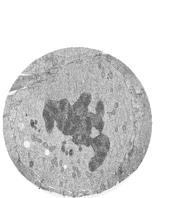 |
FIB-SEM of a dividing cell at 6.3 min after anaphase onset [3206 multi-frame micrographs composed of 1 frames each in TIFF format] | Otsuka S, Steyer AM, Schorb M, Hériché JK, Hossain MJ, Sethi S, Kueblbeck M, Schwab Y, Beck M, Ellenberg J [Pubmed: 29323269] [DOI: 10.1038/s41594-017-0001-9] |
83.2 GB | — |
| 2025-06-30 |  |
FIB-SEM Trypanosoma cruzi cell samples [multiple data sets in TIFF and PNG formats] | Slamková D., Hurník Konečná T, Sviben S | 10.8 GB | — |
| 2026-04-20 |  |
Single-particle cryo-EM data of the high-affinity choline transporter in DDM with Na+ and choline [21999 multi-frame micrographs composed of 50 frames each in TIFF format] | Vilchez-Garcia J, Jiang H, Lopez-Alonso JP, Ubarretxena-Belandia I, Tascon I | 8.1 TB | — |
| 2017-11-28 | 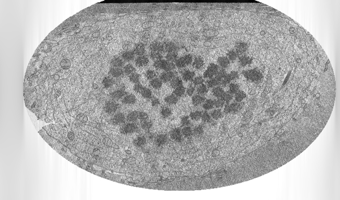 |
FIB-SEM of a dividing cell at 3.9 min after anaphase onset [2293 multi-frame micrographs composed of 1 frames each in TIFF format] | Otsuka S, Steyer AM, Schorb M, Hériché JK, Hossain MJ, Sethi S, Kueblbeck M, Schwab Y, Beck M, Ellenberg J [Pubmed: 29323269] [DOI: 10.1038/s41594-017-0001-9] |
26.8 GB | — |
| 2022-01-11 | 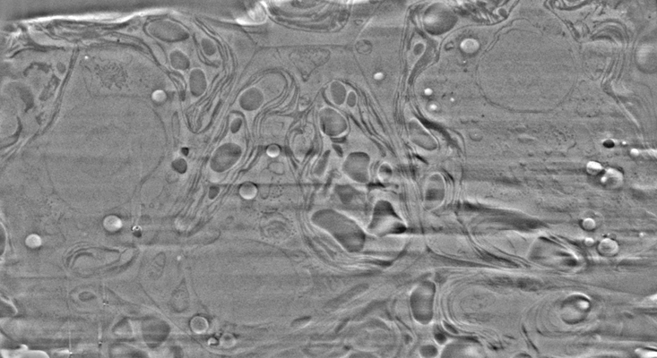 |
Cryo-FIB-SEM data on Chlamydomonas reinhardtii cells [37 micrographs in TIFF format] | Klumpe S [Pubmed: 34951584] [DOI: 10.7554/elife.70506] |
888.2 MB | — |
| 2025-06-30 |  |
FIB-SEM Nicotiana benthamiana epidermal cell - conventional acquisition [200 reconstructed volumes in TIFF format] | Czymmek KJ, Burch-Smith T, Hurník Konečná T [DOI: 10.1016/bs.mcb.2023.04.008] |
7.5 GB | — |
| 2025-11-14 |  |
Cryo-tomography of purified1.4 nm-HAN-488 labeled ribosomes from intact cells for validation of labeling method (ExoSloNano) [37 tilt series in TIFF format] | Young LN, Sherard A, Giraldez AJ, Villa E, Zhou H, Shaikh F, Hutchings J, Riggi M, Narasimhan M, Flaherty WA, Bennett E, Rosen MK | 168.9 GB | — |
| 2026-04-20 |  |
Single-particle cryo-EM data of the high-affinity choline transporter in DDM with Na+ [18599 multi-frame micrographs composed of 60 frames each in TIFF format] | Vilchez-Garcia J, Jiang H, Lopez-Alonso JP, Ubarretxena-Belandia I, Tascon I | 4.7 TB | — |
| 2026-10-02 |  |
Cryo-electron tomography of mitotic human cells (RPE1) [234 reconstructed volumes in MRC format] | Conway W, Bobe D, Fabig G, Zimyanin V, de Marco A, Redemann S [Pubmed: 41648357] [DOI: 10.64898/2026.01.16.699769] |
193.4 GB | — |
| 2017-11-30 | 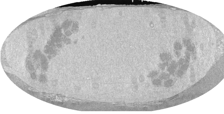 |
FIB-SEM of a dividing cell at 4.3 min after anaphase onset [1358 multi-frame micrographs composed of 1 frames each in TIFF format] | Otsuka S, Steyer AM, Schorb M, Hériché JK, Hossain MJ, Sethi S, Kueblbeck M, Schwab Y, Beck M, Ellenberg J [Pubmed: 29323269] [DOI: 10.1038/s41594-017-0001-9] |
14.0 GB | — |
| 2021-11-01 | 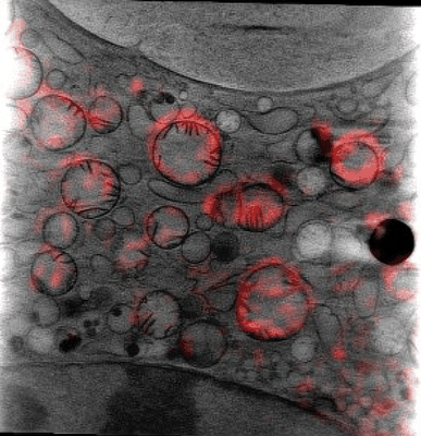 |
Cryo Soft X-ray data for tetraspeck correlation [multiple data sets in MRC format] | Groen J, Pereiro E [Pubmed: 33990802] [DOI: 10.1038/s41596-021-00522-4] |
15.1 GB | — |
| 2024-04-02 | 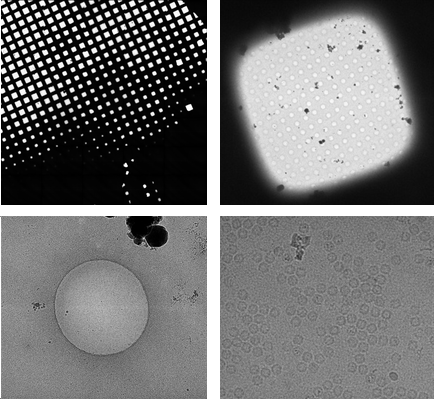 |
EMinsight: a tool to capture cryoEM microscope configuration and experimental outcomes for analysis and deposition [2605 multi-frame micrographs composed of 50 frames each in TIFF format] | Morris KL, Harrison PJ, Hatton D, Riggs S, Thiyagalingamc J | 1.2 TB | — |
| 2025-11-14 |  |
Cryo-tomography of 5 nm Nanogold labeled ribosomes from intact cells (ExoSloNano) [37 tilt series in TIFF format] | Young LN, Sherard A, Zhou H, Shaikh F, Hutchings J, Riggi M, Narasimhan M, Riggi M, Flaherty WA, Rosen MK, Giraldez AJ, Villa E [DOI: 10.1101/2024.10.12.617288] |
41.6 GB | — |
| 2017-11-30 | 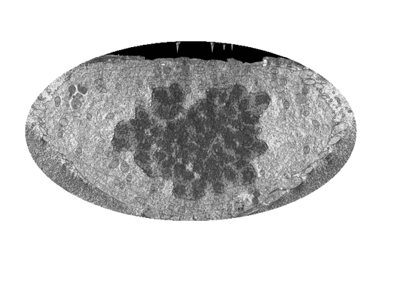 |
FIB-SEM of a dividing cell at 5.3 min after anaphase onset [1998 multi-frame micrographs composed of 1 frames each in TIFF format] | Otsuka S, Steyer AM, Schorb M, Hériché JK, Hossain MJ, Sethi S, Kueblbeck M, Schwab Y, Beck M, Ellenberg J [Pubmed: 29323269] [DOI: 10.1038/s41594-017-0001-9] |
31.2 GB | — |
| 2017-11-28 | 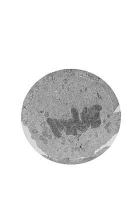 |
FIB-SEM of a dividing cell at 5.7 min after anaphase onset [2620 multi-frame micrographs composed of 1 frames each in TIFF format] | Otsuka S, Steyer AM, Schorb M, Hériché JK, Hossain MJ, Sethi S, Kueblbeck M, Schwab Y, Beck M, Ellenberg J [Pubmed: 29323269] [DOI: 10.1038/s41594-017-0001-9] |
63.0 GB | — |
| 2021-03-05 | 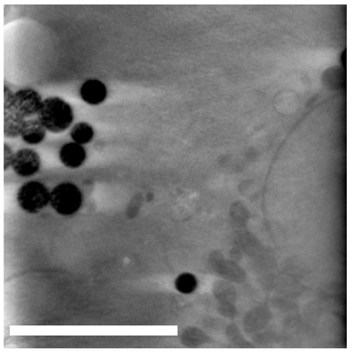 |
Dynabeads as Fiducials for SXT and SIM Correlation [700 tilt series in MRC format] | Okolo CA, Kounatidis I, Groen J, Nahas KL, Balint S, Fish T, Koronfel MA, López-Cortajarena A, Dobbie I, Pereiro E, Harkiolaki M | 1.2 GB | — |
| 2024-03-19 | 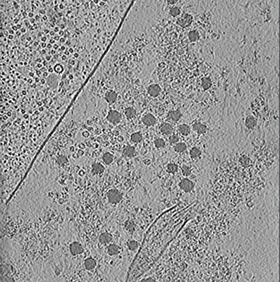 |
Unveiling the ultrastructural landscape of extracellular matrix via lift-out cryo-FIBSEM and cryo-ET [multiple data sets in TIFF and MRC formats] | Zens B., Fäßler F., Hansen J.M., Hauschild R., Datler J., Hodirnau V.V., Zheden V., Alanko J., Sixt M., Schur F.K.M. [Pubmed: 38506714] [DOI: 10.1083/jcb.202309125] |
183.0 GB | — |
