
Electron Microscopy Public Image Archive
Electron Microscopy Public Image Archive
Currently, due to a storage failure, some datasets are either inaccessible or incomplete (Marked: Incomplete dataset).
We are restoring them in ascending order of EMPIAR-ID.
If there are any entries mirrored on PDBj that are particularly urgent, we are accepting requests (email, inquiry).
The EMPIAR-PDBj team at Osaka University assists Asian EM researchers with the transfer of big EM image data to EMPIAR. Instead of sending the data directly to the EBI (UK) via the internet, hard drives can also be sent to Osaka University by postal mail or via a courier service. As an alternative, internet transfer to our server in Osaka is also available. If you would like to take advantage of our submission services, please contact us first by e-mail before sending the data to us.
| Release date | Imageset | Title | Authors and references | Size | Resolution |
|---|---|---|---|---|---|
| 2021-11-26 |  |
Cryo-EM reconstruction of heavy chain mouse apoferritin, 8 datasets testing dose rate and correlative double sampling mode for the Gatan K3 direct electron detector. [1143 multi-frame micrographs composed of 120 frames in LZW compressed TIFF format [multiple data sets in TIFF format] | Sun M, Azumaya CM, Tse E, Bulkley DP, Harrington MB, Gilbert G, Frost A, Southworth D, Verba KA, Cheng Y, Agard DA [Pubmed: 33984504] [DOI: 10.1016/j.jsb.2021.107745] |
8.5 TB | 1.655 Å |
| 2020-12-18 |  |
Cryo electron micrographs of digitonin-solubilized, amphipol-stabilized, sucrose-gradient-purified V. radiata mitochondrial membranes - mixed fraction containing CI*, CIII2 and SC III2+IV [9815 multi-frame micrographs composed of 118 frames each in TIFF format] | Maldonado M, Guo F, Letts JA [Pubmed: 33463523] [DOI: 10.7554/eLife.62047] |
6.7 TB | 3.2 - 3.9 Å |
| 2021-01-27 |  |
Cryo-EM structure of the Cas12f1-sgRNA-target DNA complex [2848 multi-frame micrographs composed of 48 frames each in TIFF format] | Takeda NS, Nakagawa R, Okazaki S, Hirano H, Kobayashi K, Kusakizako T, Nishizawa T, Yamashita K, Nishimasu H, Nureki O [Pubmed: 33333018] [DOI: 10.1016/j.molcel.2020.11.035] |
649.5 GB | 3.3 Å |
| 2020-12-04 |  |
Single-particle Cryo-EM of the Escherichia coli McrBC complex [1161 multi-frame micrographs composed of 80 frames each in TIFF format] | Niu Y, Suzuki H, Hosford CJ, Walz T, Chappie JS [Pubmed: 33219217] [DOI: 10.1038/s41467-020-19735-4] |
702.5 GB | 3.28 Å |
| 2020-12-04 |  |
Single-particle Cryo-EM of the Thermococcus gammatolerans McrBC complex [2178 multi-frame micrographs composed of 40 frames each in TIFF format] | Niu Y, Suzuki H, Hosford CJ, Walz T, Chappie JS [Pubmed: 33219217] [DOI: 10.1038/s41467-020-19735-4] |
883.3 GB | 2.44 Å |
| 2020-12-04 |  |
Single-particle Cryo-EM of the Thermococcus gammatolerans McrB AAA+ hexamer [1605 multi-frame micrographs composed of 40 frames each in TIFF format] | Niu Y, Suzuki H, Hosford CJ, Walz T, Chappie JS [Pubmed: 33219217] [DOI: 10.1038/s41467-020-19735-4] |
644.6 GB | 3.1 Å |
| 2020-12-04 | 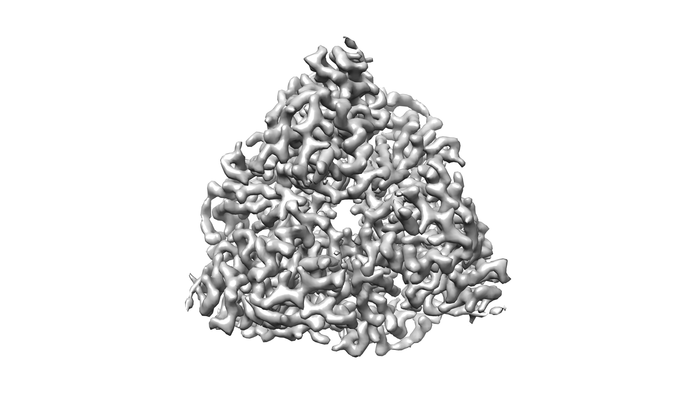 |
CryoEM map and model of Nitrite Reductase at pH 8.1 [694 multi-frame micrographs composed of 49 frames each in MRC format] | Adachi N, Yamaguchi T, Moriya T, Kawasaki M, Koiwai K, Shinoda A, Yamada Y, Yumoto F, Kohzuma T, Senda T [Pubmed: 34217801] [DOI: 10.1016/j.jsb.2021.107768] |
1.0 TB | 2.85 Å |
| 2020-12-22 | 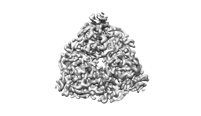 |
CryoEM map and model of Nitrite Reductase at pH 6.2 [794 multi-frame micrographs composed of 49 frames each in MRC format] | Adachi N, Yamaguchi T, Moriya T, Kawasaki M, Koiwai K, Shinoda A, Yamada Y, Yumoto F, Kohzuma T, Senda T [Pubmed: 34217801] [DOI: 10.1016/j.jsb.2021.107768] |
1.2 TB | 2.99 Å |
| 2020-12-07 | 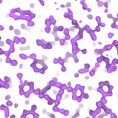 |
Krios G4 apoferritin test with K3/(slit out) SerialEM BIS 1x1x4 [4184 multi-frame micrographs composed of 76 frames each in TIFF format] | Danev R [Pubmed: 33969878] [DOI: 10.1093/jmicro/dfab016] |
1.2 TB | 1.43 Å |
| 2020-12-22 |  |
Cryo electron microscopy single particle of PIKfyve lipid kinase complex [2838 multi-frame micrographs composed of 31 frames each in TIFF format] | Lees JA, Li P, Reinisch KM [Pubmed: 33098764] [DOI: 10.1016/j.molcel.2020.10.003] |
920.4 GB | 5.25 Å |
| 2020-12-02 | 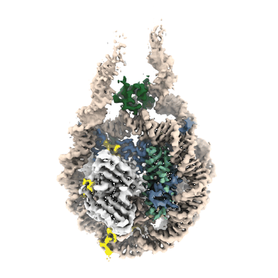 |
Single-particle cryo-EM micrographs of a chromatosome containing chimeric linker histone gH1.10-ncH1.4 [1426 multi-frame micrographs composed of 40 frames each in TIFF format] | Zhou BR [Pubmed: 33238161] [DOI: 10.1016/j.molcel.2020.10.038] |
571.7 GB | 3.03 Å |
| 2020-12-02 |  |
Single-particle cryo-EM micrographs of 197bp nucleosome with scFv [1986 multi-frame micrographs composed of 40 frames each in TIFF format] | Zhou BR [Pubmed: 33238161] [DOI: 10.1016/j.molcel.2020.10.038] |
738.9 GB | 2.85 Å |
| 2020-12-02 |  |
Single-particle cryo-EM micrographs of a chromatosome containing human linker histone H1.10 [667 multi-frame micrographs composed of 38 frames each in TIFF format] | Zhou BR [Pubmed: 33238161] [DOI: 10.1016/j.molcel.2020.10.038] |
278.2 GB | 3.12 Å |
| 2020-12-02 |  |
Single-particle cryo-EM micrographs of a chromatosome containing human linker histone H1.4 [1183 multi-frame micrographs composed of 76 frames each in TIFF format] | Zhou BR [Pubmed: 33238161] [DOI: 10.1016/j.molcel.2020.10.038] |
208.9 GB | 2.76 Å |
| 2020-12-02 |  |
Single-particle cryo-EM micrographs of a chromatosome containing human linker histone H1.0 [1069 multi-frame micrographs composed of 76 frames each in TIFF format] | Zhou BR [Pubmed: 33238161] [DOI: 10.1016/j.molcel.2020.10.038] |
349.6 GB | 2.93 Å |
| 2020-12-02 | 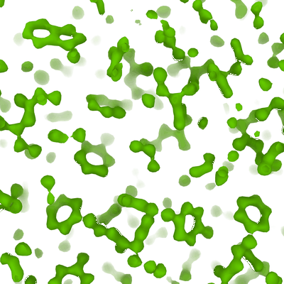 |
Krios G4 apoferritin test with K3/BioQuantum SerialEM BIS 1x1x4 [3472 multi-frame micrographs composed of 76 frames each in TIFF format] | Danev R [Pubmed: 33969878] [DOI: 10.1093/jmicro/dfab016] |
966.0 GB | 1.35 Å |
| 2021-11-16 | 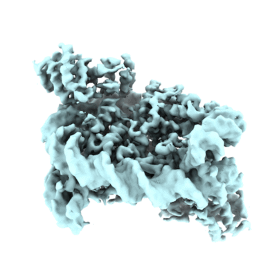 |
The cryo-EM structure of the CENP-A nucleosome in complex with the phosphorylated CENP-C:: CENP-A nucleosome in complex with CENP-C motif (655-675) and CENP-N N-terminal domain (1-211) [4630 multi-frame micrographs composed of 50 frames each in TIFF format] | Ariyoshi M, Makino F, Watanabe R, Nakagawa R, Kato T, Namba K, Arimura Y, Fujita R, Kurumizaka H, Okumura EI, Hara M, Fukagawa T [Pubmed: 33463726] [DOI: 10.15252/embj.2020105671] |
1.3 TB | 4.2 Å |
| 2021-01-22 | 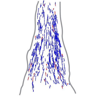 |
Actin filament structure from focal adhesions of mouse embryonic fibroblasts [7 tilt series in MRC format] | Martins B, Sorrentino S, Chung WL, Tatli M, Medalia O, Eibauer M [Pubmed: 33476550] [DOI: 10.1016/j.str.2020.12.014] |
314.1 GB | 17.0 Å |
| 2021-11-16 | 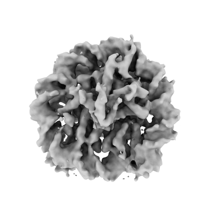 |
The cryo-EM structure of the CENP-A nucleosome in complex with the phosphorylated CENP-C:: CENP-A nucleosome in complex with phosphorylated CENP-C C-terminal domain(601-864) [6533 multi-frame micrographs composed of 50 frames each in TIFF format] | Ariyoshi M, Makino F, Watanabe R, Nakagawa R, Kato T, Namba K, Arimura Y, Fujita R, Kurumizaka H, Okumura EI, Hara M, Fukagawa T [Pubmed: 33463726] [DOI: 10.15252/embj.2020105671] |
1.4 TB | 6.8 Å |
| 2020-12-11 | 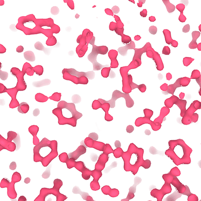 |
Krios G4 apoferritin test with Falcon 4 (no filter) EPU 1x1x4 [3540 multi-frame micrographs composed of 720 frames each in EER format] | Danev R [Pubmed: 33969878] [DOI: 10.1093/jmicro/dfab016] |
879.2 GB | 1.43 Å |
| 2020-12-09 | 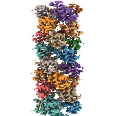 |
CARD8-CT filament [1208 multi-frame micrographs composed of 50 frames each in TIFF format] | Robert Hollingsworth L, David L, Li Y, Sharif H, Fontana P, Fu TM, Wu H [Pubmed: 33420033] [DOI: 10.1038/s41467-020-20320-y] |
1.2 TB | 3.54 Å |
| 2020-12-07 |  |
ASC-CARD:Caspase-1-CARD octamer [5377 multi-frame micrographs composed of 50 frames each in TIFF format] | Robert Hollingsworth L, David L, Li Y, Ruan J, Wu H [Pubmed: 33420033] [DOI: 10.1038/s41467-020-20320-y] |
1.2 TB | 5.0 Å |
| 2021-11-16 | 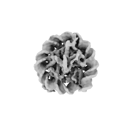 |
The cryo-EM structure of the CENP-A nucleosome in complex with the phosphorylated CENP-C: CENP-A nucleosome in complex with phosphorylated CENP-C C-terminal domain (601-864) and CENP-N N-terminal domain (1-211) [8017 multi-frame micrographs composed of 50 frames each in TIFF format] | Ariyoshi M, Makino F, Watanabe R, Nakagawa R, Kato T, Namba K, Arimura Y, Fujita R, Kurumizaka H, Okumura EI, Hara M, Fukagawa T [Pubmed: 33463726] [DOI: 10.15252/embj.2020105671] |
1.6 TB | 4.5 - 7.8 Å |
| 2020-12-04 | 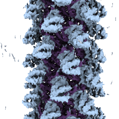 |
NLRP1-CT filament [1994 multi-frame micrographs composed of 50 frames each in TIFF format] | Robert Hollingsworth L, David L, Li Y, Wu H [Pubmed: 33420033] [DOI: 10.1038/s41467-020-20320-y] |
534.7 GB | 3.72 Å |
| 2021-04-14 |  |
Micro X ray tomography of mesoscopic placental tissue [multiple data sets in TIFF and BIG DATA VIEWER HDF5 formats] | Tun WM, Poologasundarampillai G, Bischof H, Nye G, King ONF, Basham M, Tokudome Y, Lewis RM, Johnstone ED, Brownbill P, Darrow M, Chernyavsky IL [Pubmed: 34062108] [DOI: 10.1098/rsif.2021.0140] |
50.5 GB | — |
